Publication title (প্রকাশনার শিরোনাম): Tibia and Fibula Fractures: Anatomical Levels, Clinical Definitions, Classifications, Prescription-Ready X-ray Orders, Conservative and Operative Management — টিবিয়া ও ফিবুলা ফ্র্যাকচার: অবস্থান, ধরন, সঠিক এক্স-রে পরীক্ষা ও চিকিৎসা।
Department (বিভাগ): Orthopedics / Orthopedic Trauma — অর্থোপেডিকস ও ট্রমা। Content type (ধরন): Bilingual educational medical review — বাংলা–ইংরেজি শিক্ষামূলক মেডিকেল রিভিউ। Prepared (প্রস্তুত): 29 October 2026। Status (অবস্থা): Proposed, not published — প্রস্তাবিত, প্রকাশিত নয়।
Clinical note (চিকিৎসাগত সতর্কতা): এই লেখার Imaging Orders হলো চিকিৎসকদের জন্য নমুনা Clinical Wording, কোনো রোগীর জন্য স্বয়ংক্রিয় প্রেসক্রিপশন নয়। সব পরীক্ষার ক্ষেত্রে সঠিক Side (Right/Left), Examination Findings এবং Local Radiology Protocol মেনে চলতে হবে। একই Fracture Type কখনও Conservative, কখনও Operative হতে পারে।
Details Info:- 🦴 TC(1457).(05). Orthopedics Department Case No (191)
Tibia and Fibula Fractures: Complete Clinical Classification, X-ray Investigations, Conservative Management and Operative Treatment
টিবিয়া ও ফিবুলা হাড়ের ফ্র্যাকচার: প্রতিটি প্রকারের ক্লিনিক্যাল সংজ্ঞা, সঠিক এক্স-রে পরীক্ষা, অপারেশন ছাড়া চিকিৎসা এবং অপারেশনের নির্দেশনা
LIPU MED — Evidence-Based Orthopedics Clinical Education
Publication Information — প্রকাশনা তথ্য
| Information (ইংরেজি) | বাংলা অর্থ ও বিবরণ |
|---|---|
| Total Case (TC): 1457 | মোট কেস নম্বর: ১৪৫৭ (প্রস্তাবিত) |
| Department Category Serial (DCS): 05 | বিভাগের ক্যাটাগরি সিরিয়াল: ০৫ |
| Orthopedics Case No: 191 | অর্থোপেডিকস বিভাগের কেস নম্বর: ১৯১ (প্রস্তাবিত) |
| Department: Orthopedics | বিভাগ: অস্থি ও জয়েন্ট চিকিৎসা |
| Topic: Tibia and Fibula Fractures | বিষয়: টিবিয়া ও ফিবুলা হাড়ের ফ্র্যাকচার |
| Article Type: Clinical Educational Review | চিকিৎসাবিজ্ঞানভিত্তিক শিক্ষামূলক পর্যালোচনা |
| Language: English and Bengali | ভাষা: ইংরেজি ও বাংলা |
| Patient-Specific Prescription: No | কোনো নির্দিষ্ট রোগীর প্রেসক্রিপশন নয় |
🟨 1. Introduction — ভূমিকা
1.1 Fracture — হাড় ভাঙা কী?
Clinical Definition (ক্লিনিক্যাল সংজ্ঞা):
A fracture is a partial or complete disruption of the structural continuity of a bone.
বাংলা অর্থ:
হাড়ের স্বাভাবিক গঠনগত ধারাবাহিকতা আংশিক বা সম্পূর্ণ বিচ্ছিন্ন হয়ে যাওয়াকে Fracture বা হাড় ভাঙা বলা হয়।
1.2 Tibial Fracture — টিবিয়া হাড়ের ফ্র্যাকচার
Clinical Definition:
A tibial fracture is a partial or complete break in the tibia, involving its proximal end, shaft, or distal end.
বাংলা অর্থ:
পায়ের হাঁটু থেকে গোড়ালি পর্যন্ত অবস্থিত বড় হাড় Tibia-এর উপরের, মাঝের অথবা নিচের অংশ আংশিক বা সম্পূর্ণ ভেঙে যাওয়াকে Tibial Fracture বলে।
1.3 Fibular Fracture — ফিবুলা হাড়ের ফ্র্যাকচার
Clinical Definition:
A fibular fracture is a break in the fibula that may occur independently or together with tibial and ankle injuries.
বাংলা অর্থ:
পায়ের বাইরের দিকে অবস্থিত সরু হাড় Fibula ভেঙে যাওয়াকে Fibular Fracture বলা হয়।
এটি একা অথবা Tibia ও Ankle Joint-এর অন্যান্য আঘাতের সঙ্গে হতে পারে।
Tibia ও Fibula Fracture কত প্রকার?
Fracture শ্রেণিবিন্যাসের একটি নির্দিষ্ট মোট সংখ্যা নেই।
কারণ একই Fracture-কে বিভিন্নভাবে শ্রেণিবিন্যাস করা যায়।
প্রধান শ্রেণিবিন্যাস:
- Anatomical Classification — অবস্থানভিত্তিক শ্রেণিবিন্যাস।
- Fracture Pattern Classification — হাড় ভাঙার আকৃতিভিত্তিক শ্রেণিবিন্যাস।
- Displacement Classification — ভাঙা অংশ কতটা সরে গেছে।
- Open and Closed Fracture — খোলা ও বন্ধ ফ্র্যাকচার।
- Articular Classification — জয়েন্ট জড়িত আছে কি না।
- AO/OTA Classification — আন্তর্জাতিক অস্থিভঙ্গ শ্রেণিবিন্যাস।
- Schatzker Classification — টিবিয়াল প্লাটোর ফ্র্যাকচার শ্রেণিবিন্যাস।
- Danis–Weber Classification — গোড়ালির ফিবুলা ফ্র্যাকচার শ্রেণিবিন্যাস।
- Gustilo–Anderson Classification — ওপেন ফ্র্যাকচারের শ্রেণিবিন্যাস।
- Salter–Harris Classification — শিশুদের Growth Plate Fracture-এর শ্রেণিবিন্যাস।
🟨 2. Clinical Anatomy of Tibia and Fibula — টিবিয়া ও ফিবুলার ক্লিনিক্যাল অ্যানাটমি
2.1 Tibia — টিবিয়া
Tibia হলো পায়ের নিচের অংশের প্রধান ওজনবাহী হাড়।
এর প্রধান তিনটি অংশ:
| English Term | বাংলা অর্থ |
|---|---|
| Proximal Tibia | টিবিয়ার উপরের অংশ, হাঁটুর কাছে |
| Tibial Shaft | টিবিয়ার মাঝের লম্বা অংশ |
| Distal Tibia | টিবিয়ার নিচের অংশ, গোড়ালির কাছে |
Proximal Tibia-এর গুরুত্বপূর্ণ অংশ
- Tibial Plateau — হাঁটুর নিচের জয়েন্টের সমতল পৃষ্ঠ।
- Medial Tibial Condyle — টিবিয়ার উপরের ভেতরের অংশ।
- Lateral Tibial Condyle — টিবিয়ার উপরের বাইরের অংশ।
- Tibial Spine — টিবিয়ার উপরের মধ্যবর্তী উঁচু অংশ।
- Tibial Tuberosity — হাঁটুর নিচে Patellar Tendon সংযুক্ত হওয়ার স্থান।
- Metaphysis — হাড়ের প্রান্ত ও Shaft-এর মধ্যবর্তী অঞ্চল।
Distal Tibia-এর গুরুত্বপূর্ণ অংশ
- Tibial Plafond — গোড়ালির জয়েন্টের ওপরের ওজনবাহী পৃষ্ঠ।
- Medial Malleolus — গোড়ালির ভেতরের হাড়ের উঁচু অংশ।
- Posterior Malleolus — টিবিয়ার নিচের পেছনের অংশ।
2.2 Fibula — ফিবুলা
Fibula হলো পায়ের বাইরের দিকে অবস্থিত অপেক্ষাকৃত সরু হাড়।
এর অংশগুলো:
| English Term | বাংলা অর্থ |
|---|---|
| Fibular Head | ফিবুলার উপরের মাথা |
| Fibular Neck | ফিবুলার মাথার নিচের সরু অংশ |
| Fibular Shaft | ফিবুলার মাঝের লম্বা অংশ |
| Lateral Malleolus | গোড়ালির বাইরের হাড়ের উঁচু অংশ |
Clinical Importance — চিকিৎসাগত গুরুত্ব:
Fibular Neck-এর কাছে Common Fibular Nerve বা Common Peroneal Nerve অবস্থান করে।
এই অঞ্চলে Injury হলে Foot Drop হতে পারে।
Foot Drop — পায়ের পাতা ওপরের দিকে তুলতে অসুবিধা হওয়া।
তাই Proximal Fibular Fracture-এর ক্ষেত্রে Nerve Function পরীক্ষা করা জরুরি।
🟨 3. Anatomical Classification and Prescription-Ready X-ray Orders
অবস্থান অনুযায়ী Fracture ও প্রেসক্রিপশনে সঠিক X-ray লেখার নিয়ম
Clinical Prescription Format — প্রেসক্রিপশন লেখার নিয়ম
Investigation Name + Side + Anatomical Region + Required Views
বাংলা অর্থ:
পরীক্ষার নাম + ডান/বাম দিক + শরীরের নির্দিষ্ট অংশ + প্রয়োজনীয় এক্স-রে ভিউ।
Right এবং Left লেখার নিয়ম
| English | বাংলা |
|---|---|
| Right / Rt. | ডান |
| Left / Lt. | বাম |
| Bilateral | উভয় পাশ |
| AP View | Anteroposterior — সামনে থেকে পেছনের অভিমুখে নেওয়া এক্স-রে |
| Lateral View | পাশ থেকে নেওয়া এক্স-রে |
| Mortise View | গোড়ালির জয়েন্টের Alignment দেখার বিশেষ এক্স-রে ভিউ |
| Oblique View | তির্যক কোণ থেকে নেওয়া এক্স-রে |
3.1 Prescription-Ready X-ray Table
নিচের উদাহরণগুলোতে Right Side ব্যবহার করা হয়েছে। রোগীর আঘাত বাম পায়ে হলে Right-এর পরিবর্তে Left লিখতে হবে।
| Suspected Fracture — সম্ভাব্য ফ্র্যাকচার | Prescription-Ready Investigation |
|---|---|
| Tibial Plateau Fracture — হাঁটুর টিবিয়াল প্লাটো ভাঙা | X-ray Right Knee Joint — AP and Lateral Views |
| Tibial Spine Fracture — টিবিয়াল স্পাইন ভাঙা | X-ray Right Knee Joint — AP and Lateral Views |
| Tibial Tuberosity Fracture — টিবিয়াল টিউবেরোসিটি ভাঙা | X-ray Right Knee Joint — AP and Lateral Views |
| Proximal Tibial Metaphyseal Fracture — টিবিয়ার উপরের মেটাফাইসিস ভাঙা | X-ray Right Knee Joint — AP and Lateral Views; include adjacent proximal tibia |
| Tibial Shaft Fracture — টিবিয়ার মাঝের অংশ ভাঙা | X-ray Right Leg (Tibia and Fibula) — AP and Lateral Views, including Knee and Ankle Joints |
| Fibular Shaft Fracture — ফিবুলার মাঝের অংশ ভাঙা | X-ray Right Leg (Tibia and Fibula) — AP and Lateral Views, including Knee and Ankle Joints |
| Proximal Fibula Fracture — ফিবুলার উপরের অংশ ভাঙা | X-ray Right Knee Joint — AP and Lateral Views |
| Distal Tibia Fracture — টিবিয়ার নিচের অংশ ভাঙা | X-ray Right Leg (Tibia and Fibula) — AP and Lateral Views; dedicated Ankle Views if indicated |
| Pilon Fracture — গোড়ালির জয়েন্টের টিবিয়াল প্লাফন্ড ভাঙা | X-ray Right Ankle Joint — AP, Lateral and Mortise Views |
| Medial Malleolus Fracture — গোড়ালির ভেতরের অংশ ভাঙা | X-ray Right Ankle Joint — AP, Lateral and Mortise Views |
| Posterior Malleolus Fracture — গোড়ালির পেছনের অংশ ভাঙা | X-ray Right Ankle Joint — AP, Lateral and Mortise Views |
| Lateral Malleolus Fracture — গোড়ালির বাইরের অংশ ভাঙা | X-ray Right Ankle Joint — AP, Lateral and Mortise Views |
| Weber A, B or C Injury — ফিবুলার গোড়ালিসংলগ্ন ফ্র্যাকচার | X-ray Right Ankle Joint — AP, Lateral and Mortise Views |
| Suspected Maisonneuve Injury — গোড়ালি ও ফিবুলার উপরের অংশের যৌথ আঘাত | X-ray Right Ankle Joint — AP, Lateral and Mortise Views + X-ray Right Leg (Tibia and Fibula) — AP and Lateral Views |
Important Clinical Note — গুরুত্বপূর্ণ ক্লিনিক্যাল তথ্য
Tibial Shaft Fracture-এর ক্ষেত্রে Complete Tibia–Fibula Radiographs নেওয়ার সময় Knee এবং Ankle Joint অন্তর্ভুক্ত করা গুরুত্বপূর্ণ।
তবে নির্দিষ্ট Knee বা Ankle Joint-এর অতিরিক্ত Injury সন্দেহ হলে Dedicated Joint Radiographs আলাদাভাবে প্রয়োজন হতে পারে।
Advanced Imaging — অতিরিক্ত পরীক্ষা
CT Scan Without IV Contrast — শিরায় কনট্রাস্ট না দিয়ে সিটি স্ক্যান।
Complex Articular Fracture-এর গঠন, Depression ও Displacement ভালোভাবে বোঝার জন্য ব্যবহৃত হয়।
MRI Without IV Contrast — শিরায় কনট্রাস্ট না দিয়ে এমআরআই।
Ligament, Meniscus, Occult Fracture অথবা Stress Fracture-এর নির্বাচিত মূল্যায়নে ব্যবহৃত হয়।
🟨 4. Proximal Tibia Fractures — টিবিয়ার উপরের অংশের ফ্র্যাকচার
4.1 Tibial Plateau Fracture — টিবিয়াল প্লাটো ফ্র্যাকচার
Clinical Definition — ক্লিনিক্যাল সংজ্ঞা
A tibial plateau fracture is an intra-articular fracture involving the proximal articular surface of the tibia.
বাংলা অর্থ:
হাঁটুর জয়েন্ট গঠনকারী Tibia-এর উপরের Articular Surface ভেঙে যাওয়াকে Tibial Plateau Fracture বলা হয়।
Clinical Features — লক্ষণ
- Knee Pain — হাঁটুতে ব্যথা।
- Knee Swelling — হাঁটু ফুলে যাওয়া।
- Joint Effusion — জয়েন্টে অতিরিক্ত তরল জমা।
- Hemarthrosis — জয়েন্টের ভেতরে রক্ত জমা।
- Difficulty in Weight Bearing — পায়ে ভর দিতে অসুবিধা।
- Knee Instability — হাঁটুর স্থিতিশীলতা কমে যাওয়া।
Prescription-Ready X-ray
X-ray Right Knee Joint — AP and Lateral Views.
Clinical Indication: Suspected Tibial Plateau Fracture.
বাংলা অর্থ: টিবিয়াল প্লাটো ফ্র্যাকচারের সন্দেহে ডান হাঁটুর সামনের ও পাশের এক্স-রে।
Additional Investigation
CT Scan Right Knee Joint Without IV Contrast.
বাংলা অর্থ: ডান হাঁটুর জয়েন্টের কনট্রাস্টবিহীন সিটি স্ক্যান।
এটি Complex Fracture, Articular Depression ও Operative Planning-এর জন্য প্রয়োজন হতে পারে।
Schatzker Classification — ছয়টি প্রকার
Type I — Lateral Plateau Split Fracture
Clinical Definition:
A fracture characterized by splitting of the lateral tibial plateau without major articular depression.
বাংলা অর্থ:
টিবিয়াল প্লাটোর বাইরের অংশে ফাটল সৃষ্টি হয়ে আলাদা Fragment তৈরি হয়, কিন্তু Joint Surface উল্লেখযোগ্যভাবে দেবে যায় না।
X-ray Findings — এক্স-রে-তে যা দেখা যায়:
Lateral Tibial Plateau-তে Split Fracture Line।
বাংলা: টিবিয়াল প্লাটোর বাইরের অংশে বিভক্ত ফ্র্যাকচার রেখা।
Investigation:
X-ray Right Knee Joint — AP and Lateral Views।
CT Right Knee Without IV Contrast — প্রয়োজন অনুযায়ী।
Conservative Treatment — অপারেশন ছাড়া চিকিৎসা:
Stable ও Minimally Displaced Fracture হলে:
- Hinged Knee Brace — হাঁটুর নড়াচড়া নিয়ন্ত্রণকারী ব্রেস।
- Protected Weight Bearing — সীমিত বা নিয়ন্ত্রিত ভর দেওয়া।
- Serial X-ray — নির্দিষ্ট সময় পরপর এক্স-রে।
- Supervised Knee Mobilization — চিকিৎসকের নির্দেশে হাঁটু নাড়ানোর ব্যায়াম।
OT Indication — অপারেশনের কারণ:
Significant Displacement, Joint Incongruity অথবা Knee Instability।
Operative Treatment — অস্ত্রোপচার:
ORIF with Lag Screws or Plate Fixation।
বাংলা: ভাঙা হাড় সঠিক অবস্থানে এনে স্ক্রু অথবা প্লেট দিয়ে স্থির করা।
Type II — Lateral Split-Depression Fracture
Clinical Definition:
A fracture involving a lateral plateau split together with depression of the articular surface.
বাংলা অর্থ:
টিবিয়াল প্লাটোর বাইরের অংশ ভেঙে আলাদা হওয়ার পাশাপাশি জয়েন্টের পৃষ্ঠ নিচে দেবে যাওয়া।
X-ray Findings:
Lateral Plateau Split and Articular Depression।
বাংলা: জয়েন্টের বাইরের অংশে ফাটল ও নিচের দিকে দেবে যাওয়া।
Investigation:
X-ray Right Knee Joint — AP and Lateral Views।
CT Right Knee Without IV Contrast।
Conservative Treatment:
Minimal Depression, Stable Joint এবং Acceptable Alignment থাকলে নির্বাচিত ক্ষেত্রে Brace ও Restricted Weight Bearing।
OT Indication:
Significant Articular Depression, Displacement অথবা Knee Instability।
Operative Treatment:
Articular Surface Elevation and ORIF with Buttress Plate।
বাংলা: দেবে যাওয়া জয়েন্টের পৃষ্ঠ উঁচু করে প্লেট ও স্ক্রুর সাহায্যে স্থির করা।
প্রয়োজন অনুযায়ী Bone Graft ব্যবহার হতে পারে।
Type III — Pure Lateral Plateau Depression Fracture
Clinical Definition:
A fracture characterized by depression of the lateral tibial plateau articular surface without a major split fragment.
বাংলা অর্থ:
টিবিয়াল প্লাটোর বাইরের জয়েন্টের পৃষ্ঠ নিচে দেবে যায়, কিন্তু বড় কোনো Split Fragment থাকে না।
X-ray Findings:
Depressed Lateral Articular Surface।
বাংলা: টিবিয়ার বাইরের জয়েন্টের পৃষ্ঠ নিচের দিকে বসে যাওয়া।
Investigation:
X-ray Right Knee Joint — AP and Lateral Views।
CT Right Knee Without IV Contrast।
Conservative Treatment:
Minimal Depression ও Stable Knee হলে।
OT Indication:
Significant Depression, Joint Incongruity অথবা Instability।
Operative Treatment:
Articular Elevation, Bone Defect Support and Internal Fixation।
বাংলা: দেবে যাওয়া হাড়ের অংশ ওপরে তুলে প্রয়োজনীয় সাপোর্ট দিয়ে স্থির করা।
Type IV — Medial Tibial Plateau Fracture
Clinical Definition:
An intra-articular fracture involving the medial tibial plateau.
বাংলা অর্থ:
হাঁটুর ভেতরের Tibial Plateau ভেঙে যাওয়া।
X-ray Findings:
Medial Plateau Fracture Line।
বাংলা: টিবিয়াল প্লাটোর ভেতরের অংশে ফ্র্যাকচার রেখা।
Investigation:
X-ray Right Knee Joint — AP and Lateral Views।
CT Right Knee Without IV Contrast।
Conservative Treatment:
Stable, Undisplaced Fracture-এর নির্বাচিত ক্ষেত্রে।
OT Indication:
Displacement, Varus Instability অথবা Joint Incongruity।
Operative Treatment:
ORIF with Medial or Posteromedial Buttress Plate।
বাংলা: টিবিয়ার ভেতরের অথবা পেছনের-ভেতরের দিকে প্লেট দিয়ে হাড় স্থির করা।
Type V — Bicondylar Tibial Plateau Fracture
Clinical Definition:
A tibial plateau fracture involving both medial and lateral condyles.
বাংলা অর্থ:
টিবিয়াল প্লাটোর ভেতরের এবং বাইরের উভয় অংশ একসঙ্গে ভেঙে যাওয়া।
X-ray Findings:
Medial and Lateral Plateau Fractures।
বাংলা: টিবিয়াল প্লাটোর উভয় অংশে ফ্র্যাকচার।
Investigation:
X-ray Right Knee Joint — AP and Lateral Views।
CT Right Knee Without IV Contrast।
Conservative Treatment:
শুধু নির্বাচিত Stable ও Minimally Displaced Injury-তে বিবেচনা করা হয়।
OT Indication:
Displaced Bicondylar Fracture, Articular Incongruity অথবা Instability।
Operative Treatment:
ORIF with Appropriate Plate and Screw Fixation।
বাংলা: প্রয়োজন অনুযায়ী এক বা একাধিক প্লেট ও স্ক্রুর সাহায্যে হাড়ের অংশ স্থির করা।
Soft Tissue Swelling গুরুতর হলে Staged Fixation প্রয়োজন হতে পারে।
Type VI — Metaphyseal–Diaphyseal Dissociation
Clinical Definition:
A tibial plateau fracture in which the articular segment is separated from the tibial shaft.
বাংলা অর্থ:
টিবিয়ার ওপরের জয়েন্ট গঠনকারী অংশ এবং নিচের Shaft-এর মধ্যে হাড়ের ধারাবাহিক সংযোগ বিচ্ছিন্ন হয়ে যাওয়া।
X-ray Findings:
Separation of the Articular Segment from the Tibial Shaft।
বাংলা: জয়েন্টের অংশ ও টিবিয়ার Shaft-এর মধ্যে বিচ্ছিন্নতা।
Investigation:
X-ray Right Knee Joint — AP and Lateral Views।
X-ray Right Leg (Tibia and Fibula) — AP and Lateral Views, if required।
CT Right Knee Without IV Contrast।
Conservative Treatment:
খুব সীমিত Selected Stable Case-এ।
OT Indication:
Displaced বা Unstable Metaphyseal–Diaphyseal Dissociation।
Operative Treatment:
Temporary External Fixation followed by Definitive ORIF, if indicated।
বাংলা: প্রথমে বাহ্যিক ফিক্সেটর দিয়ে হাড় স্থির করে পরবর্তীতে প্লেট-স্ক্রু দিয়ে স্থায়ীভাবে স্থির করা।
4.2 Tibial Spine Fracture — টিবিয়াল স্পাইন ফ্র্যাকচার
Clinical Definition:
An avulsion fracture of the tibial intercondylar eminence, commonly involving the anterior cruciate ligament attachment.
বাংলা অর্থ:
হাঁটুর ACL Ligament সংযুক্ত থাকা টিবিয়ার মধ্যবর্তী উঁচু অংশ টানের কারণে ভেঙে আলাদা হয়ে যাওয়া।
Prescription X-ray:
X-ray Right Knee Joint — AP and Lateral Views।
X-ray Findings:
Avulsed Bone Fragment at the Tibial Eminence।
বাংলা: টিবিয়াল এমিনেন্সে হাড়ের বিচ্ছিন্ন টুকরা।
Conservative Treatment:
Nondisplaced বা Stable Reduced Fracture-এ Immobilization।
OT Indication:
Displaced, Irreducible অথবা Entrapped Fragment।
Operation:
Arthroscopic or Open Reduction and Fixation।
বাংলা: আর্থ্রোস্কপি বা ওপেন পদ্ধতিতে হাড়ের টুকরা সঠিক অবস্থানে এনে স্থির করা।
4.3 Tibial Tuberosity Fracture — টিবিয়াল টিউবেরোসিটি ফ্র্যাকচার
Clinical Definition:
An avulsion fracture involving the tibial tuberosity at the insertion of the patellar tendon.
বাংলা অর্থ:
Patellar Tendon-এর টানের কারণে টিবিয়ার সামনের উঁচু অংশ ভেঙে যাওয়া।
Prescription X-ray:
X-ray Right Knee Joint — AP and Lateral Views।
X-ray Findings:
Avulsion Fragment at the Tibial Tuberosity।
বাংলা: টিবিয়াল টিউবেরোসিটিতে বিচ্ছিন্ন হাড়ের অংশ।
Conservative Treatment:
Nondisplaced ও Extensor Mechanism Intact থাকলে Knee Immobilization।
OT Indication:
Significant Displacement অথবা Extensor Mechanism Dysfunction।
Operation:
ORIF with Screw Fixation।
বাংলা: স্ক্রুর সাহায্যে ভাঙা অংশ স্থির করা।
4.4 Proximal Tibial Metaphyseal Fracture — টিবিয়ার উপরের মেটাফাইসিসের ফ্র্যাকচার
Clinical Definition:
A fracture involving the proximal tibial metaphysis below the tibial plateau.
বাংলা অর্থ:
টিবিয়াল প্লাটোর নিচের Metaphysis অংশে ফ্র্যাকচার হওয়া।
Prescription X-ray:
X-ray Right Knee Joint — AP and Lateral Views, including Proximal Tibia।
Conservative Treatment:
Stable এবং Acceptably Aligned Fracture-এ Immobilization।
OT Indication:
Significant Displacement, Instability অথবা Unacceptable Angulation।
Operation:
Plate Fixation, Selected Intramedullary Nailing or External Fixation।
বাংলা: ফ্র্যাকচারের গঠন অনুযায়ী প্লেট, নেইল অথবা বাহ্যিক ফিক্সেটর ব্যবহার।
🟨 5. Tibial Shaft Fracture — টিবিয়ার মাঝের অংশের ফ্র্যাকচার
Clinical Definition — ক্লিনিক্যাল সংজ্ঞা
A tibial shaft fracture is a fracture involving the diaphyseal segment of the tibia.
বাংলা অর্থ:
টিবিয়া হাড়ের মাঝের লম্বা Diaphyseal অংশ ভেঙে যাওয়াকে Tibial Shaft Fracture বলে।
Anatomical Location
| English | বাংলা |
|---|---|
| Proximal Third | উপরের এক-তৃতীয়াংশ |
| Middle Third | মাঝের এক-তৃতীয়াংশ |
| Distal Third | নিচের এক-তৃতীয়াংশ |
Prescription-Ready Investigation
X-ray Right Leg (Tibia and Fibula) — AP and Lateral Views, including Knee and Ankle Joints.
বাংলা অর্থ:
ডান পায়ের টিবিয়া ও ফিবুলার সামনের এবং পাশের এক্স-রে, যাতে হাঁটু ও গোড়ালির জয়েন্ট অন্তর্ভুক্ত থাকে।
AO/OTA Classification — Tibial Shaft
Type A — Simple Fracture
বাংলা: অপেক্ষাকৃত সরল ফ্র্যাকচার।
Type B — Wedge Fracture
বাংলা: ভাঙা হাড়ের মাঝখানে অতিরিক্ত Wedge আকৃতির অংশ থাকা।
Type C — Multifragmentary Fracture
বাংলা: হাড় একাধিক অংশে ভেঙে যাওয়া।
5.1 AO/OTA 42-A1 — Simple Spiral Fracture
Clinical Definition:
A simple fracture in which the fracture line follows a helical course around the long axis of the tibia.
বাংলা অর্থ:
টিবিয়া হাড়ের চারপাশে প্যাঁচানো রেখা বরাবর ফ্র্যাকচার সৃষ্টি হওয়া।
X-ray Findings:
Spiral or Helical Fracture Line।
বাংলা: প্যাঁচানো আকৃতির ফ্র্যাকচার রেখা।
Prescription X-ray:
X-ray Right Leg (Tibia and Fibula) — AP and Lateral Views, including Knee and Ankle Joints।
Conservative Treatment:
Stable, Minimally Displaced অথবা Acceptably Reduced Fracture-এ Cast Immobilization।
OT Indication:
Unstable Fracture, Malrotation, Significant Displacement বা Reduction ধরে রাখা সম্ভব না হলে।
Operative Treatment:
Intramedullary Interlocking Nailing।
বাংলা: হাড়ের ভেতরের কেন্দ্রীয় নালিতে ধাতব নেইল প্রবেশ করিয়ে হাড় স্থির করা।
নির্বাচিত ক্ষেত্রে Plate Fixation করা হয়।
5.2 AO/OTA 42-A2 — Simple Oblique Fracture
Clinical Definition:
A fracture with a single oblique fracture line across the tibial shaft.
বাংলা অর্থ:
টিবিয়া হাড়ের মাঝের অংশ তির্যকভাবে ভেঙে যাওয়া।
X-ray Findings:
Oblique Fracture Line।
বাংলা: তির্যক ফ্র্যাকচার রেখা।
Prescription X-ray:
X-ray Right Leg (Tibia and Fibula) — AP and Lateral Views।
Conservative Treatment:
Stable, Acceptably Aligned Fracture-এ Cast।
OT Indication:
Unstable Displacement, Shortening অথবা Malalignment।
Operative Treatment:
IM Interlocking Nail অথবা Selected Plate Fixation।
5.3 AO/OTA 42-A3 — Simple Transverse Fracture
Clinical Definition:
A fracture with a line approximately perpendicular to the long axis of the tibia.
বাংলা অর্থ:
টিবিয়া হাড় তার লম্বা অক্ষের প্রায় সমকোণে আড়াআড়িভাবে ভেঙে যাওয়া।
X-ray Findings:
Transverse Fracture Line।
বাংলা: আড়াআড়ি ফ্র্যাকচার রেখা।
Prescription X-ray:
X-ray Right Leg (Tibia and Fibula) — AP and Lateral Views।
Conservative Treatment:
Stable, Nondisplaced অথবা Acceptably Reduced Fracture।
OT Indication:
Significant Displacement বা Unacceptable Alignment।
Operative Treatment:
IM Interlocking Nailing অথবা Selected Compression Plating।
5.4 AO/OTA 42-B2 — Intact Wedge Fracture
Clinical Definition:
A fracture with an intact wedge fragment between the two main bone fragments.
বাংলা অর্থ:
হাড়ের দুটি প্রধান ভাঙা অংশের মাঝখানে একটি অক্ষত Wedge আকৃতির হাড়ের টুকরা থাকা।
X-ray Findings:
Intact Wedge or Butterfly Fragment।
বাংলা: মাঝখানে ত্রিভুজাকার বা প্রজাপতির ডানার মতো হাড়ের অংশ।
Prescription X-ray:
X-ray Right Leg (Tibia and Fibula) — AP and Lateral Views।
Conservative Treatment:
Stable ও Minimally Displaced Fracture।
OT Indication:
Instability অথবা Significant Displacement।
Operation:
IM Nail অথবা Bridge Plating।
বাংলা: ভাঙা অংশের Alignment বজায় রাখতে নেইল অথবা প্লেট ব্যবহার।
5.5 AO/OTA 42-B3 — Fragmentary Wedge Fracture
Clinical Definition:
A wedge fracture in which the intermediate wedge component is fragmented.
বাংলা অর্থ:
হাড়ের প্রধান দুটি অংশের মাঝখানে থাকা Wedge Segment একাধিক টুকরায় ভেঙে যাওয়া।
X-ray Findings:
Fragmented Wedge Components।
বাংলা: মাঝখানের Wedge অংশ একাধিক Fragment-এ বিভক্ত।
Prescription X-ray:
X-ray Right Leg (Tibia and Fibula) — AP and Lateral Views।
Conservative Treatment:
Selected Stable Fracture।
OT Indication:
Unstable অথবা Significantly Displaced Fracture।
Operation:
IM Nail অথবা Bridge Plate Fixation।
5.6 AO/OTA 42-C2 — Intact Segmental Fracture
Clinical Definition:
A fracture occurring at two distinct levels, producing an intact intermediate bone segment.
বাংলা অর্থ:
একই হাড় দুটি আলাদা স্থানে ভেঙে মাঝখানে একটি অক্ষত Bone Segment তৈরি হওয়া।
X-ray Findings:
Two Distinct Fracture Levels।
বাংলা: একই হাড়ে দুটি আলাদা ফ্র্যাকচার স্থান।
Prescription X-ray:
X-ray Right Leg (Tibia and Fibula) — AP and Lateral Views, including Knee and Ankle Joints।
Conservative Treatment:
খুব নির্বাচিত Stable ও Acceptably Aligned Case-এ।
OT Indication:
Displacement, Instability অথবা Alignment ধরে রাখতে না পারা।
Operation:
IM Interlocking Nail, Bridge Plating বা External Fixation।
5.7 AO/OTA 42-C3 — Fragmentary Segmental Fracture
Clinical Definition:
A multifragmentary segmental fracture in which the intermediate segment is itself fragmented.
বাংলা অর্থ:
হাড়ের দুটি আলাদা স্তরে ফ্র্যাকচার হওয়ার পাশাপাশি মাঝের Bone Segment-ও একাধিক টুকরায় ভেঙে যাওয়া।
X-ray Findings:
Multiple Fragmented Segmental Components।
বাংলা: মাঝের Segment-সহ হাড়ের বিভিন্ন অংশ একাধিক টুকরায় ভাঙা।
Prescription X-ray:
X-ray Right Leg (Tibia and Fibula) — AP and Lateral Views।
Conservative Treatment:
খুব সীমিত Selected Case-এ।
OT Indication:
Unstable, Displaced অথবা Complex Fracture।
Operation:
IM Nail, Bridge Plating অথবা External Fixation।
গুরুতর Soft Tissue Injury থাকলে Staged Surgical Management প্রয়োজন হতে পারে।
🩷 6. Distal Tibia Fractures — টিবিয়ার নিচের অংশের ফ্র্যাকচার
6.1 Distal Tibial Metaphyseal Fracture
Clinical Definition:
A fracture involving the distal metaphyseal region of the tibia.
বাংলা অর্থ:
গোড়ালির কাছাকাছি টিবিয়া হাড়ের নিচের Metaphyseal অংশ ভেঙে যাওয়া।
Prescription X-ray:
X-ray Right Leg (Tibia and Fibula) — AP and Lateral Views।
Ankle-এর কাছে Injury থাকলে:
X-ray Right Ankle Joint — AP, Lateral and Mortise Views।
Conservative Treatment:
Stable ও Minimally Displaced Fracture-এ Cast।
OT Indication:
Instability, Significant Displacement অথবা Unacceptable Alignment।
Operation:
IM Nail, Plate Fixation অথবা Selected External Fixation।
6.2 Tibial Pilon Fracture — টিবিয়াল পাইলন ফ্র্যাকচার
Clinical Definition:
A pilon fracture is an intra-articular fracture involving the weight-bearing surface of the distal tibial plafond.
বাংলা অর্থ:
গোড়ালির জয়েন্টের ওপরের প্রধান ওজনবাহী টিবিয়াল পৃষ্ঠ ভেঙে যাওয়াকে Pilon Fracture বলে।
X-ray Findings:
Articular Fracture, Depression, Displacement and Possible Comminution।
বাংলা: জয়েন্টের পৃষ্ঠ ভাঙা, নিচে দেবে যাওয়া, সরে যাওয়া এবং একাধিক টুকরা হওয়া।
Prescription X-ray
X-ray Right Ankle Joint — AP, Lateral and Mortise Views.
Additional Investigation
CT Scan Right Ankle Joint Without IV Contrast.
Clinical Indication: Suspected or Confirmed Tibial Pilon Fracture; Fracture Mapping and Operative Planning।
Conservative Treatment:
Stable, Nondisplaced বা Minimally Displaced নির্বাচিত Fracture।
OT Indication:
Displaced Articular Fracture, Joint Incongruity অথবা Unstable Injury।
Operation:
Temporary External Fixation followed by Definitive ORIF, if required।
বাংলা: প্রথমে বাহ্যিক ফিক্সেটর দিয়ে হাড় স্থির করা এবং Soft Tissue-এর অবস্থা উপযুক্ত হলে পরবর্তী সময়ে প্লেট-স্ক্রু দিয়ে স্থায়ীভাবে স্থির করা।
6.3 AO/OTA 43-A — Extra-Articular Distal Tibia Fracture
Definition:
A distal tibial fracture without involvement of the articular surface.
বাংলা অর্থ:
টিবিয়ার নিচের অংশ ভেঙেছে, কিন্তু গোড়ালির জয়েন্টের পৃষ্ঠ জড়িত নয়।
X-ray:
X-ray Right Ankle Joint — AP, Lateral and Mortise Views।
প্রয়োজন অনুযায়ী Tibia–Fibula AP and Lateral।
Conservative Treatment:
Stable ও Minimally Displaced Fracture।
Operation:
Unstable বা Displaced হলে Appropriate Fixation।
6.4 AO/OTA 43-B — Partial Articular Fracture
Definition:
A fracture involving part of the articular surface while another portion remains attached to the shaft.
বাংলা অর্থ:
জয়েন্টের পৃষ্ঠের কিছু অংশ ভেঙে বিচ্ছিন্ন হলেও অন্য অংশ Shaft-এর সঙ্গে যুক্ত থাকে।
X-ray:
X-ray Right Ankle Joint — AP, Lateral and Mortise Views।
Additional Imaging:
CT Right Ankle Without IV Contrast।
Conservative Treatment:
Selected Stable, Minimally Displaced Fracture।
Operation:
Significant Articular Displacement বা Instability থাকলে ORIF।
6.5 AO/OTA 43-C — Complete Articular Fracture
Definition:
A fracture in which the entire articular segment is separated from the tibial shaft.
বাংলা অর্থ:
টিবিয়ার নিচের সম্পূর্ণ জয়েন্ট গঠনকারী অংশ Shaft থেকে বিচ্ছিন্ন হয়ে যাওয়া।
X-ray:
X-ray Right Ankle Joint — AP, Lateral and Mortise Views।
Additional Imaging:
CT Right Ankle Without IV Contrast।
Conservative Treatment:
বিরল Selected Stable Case-এ।
Operation:
Displaced/Unstable Fracture-এ সাধারণত Surgical Reconstruction and Fixation।
6.6 Medial Malleolus Fracture — গোড়ালির ভেতরের হাড়ের ফ্র্যাকচার
Clinical Definition:
A fracture involving the medial malleolus of the distal tibia.
বাংলা অর্থ:
গোড়ালির ভেতরের দিকে টিবিয়ার উঁচু হাড়ের অংশ ভেঙে যাওয়া।
Prescription X-ray:
X-ray Right Ankle Joint — AP, Lateral and Mortise Views।
Conservative Treatment:
Stable, Nondisplaced Fracture।
OT Indication:
Displacement অথবা Ankle Instability।
Operation:
ORIF with Screws or Appropriate Plate Fixation।
6.7 Posterior Malleolus Fracture — গোড়ালির পেছনের হাড়ের ফ্র্যাকচার
Clinical Definition:
A fracture involving the posterior articular margin of the distal tibia.
বাংলা অর্থ:
গোড়ালির জয়েন্ট গঠনকারী টিবিয়ার নিচের পেছনের অংশ ভেঙে যাওয়া।
Prescription X-ray:
X-ray Right Ankle Joint — AP, Lateral and Mortise Views।
Additional Investigation:
CT Right Ankle Without IV Contrast।
Conservative Treatment:
Small, Stable ও Nondisplaced Fracture।
OT Indication:
Joint Incongruity, Instability অথবা Significant Displacement।
Operation:
Posterior Malleolar Fixation with Screw or Buttress Plate।
🟪 7. Fibula Fractures — ফিবুলা হাড়ের বিভিন্ন ফ্র্যাকচার
7.1 Proximal Fibular Fracture — ফিবুলার উপরের অংশ ভাঙা
Clinical Definition:
A fracture involving the fibular head or neck.
বাংলা অর্থ:
ফিবুলার ওপরের Head অথবা Neck অংশ ভেঙে যাওয়া।
Prescription X-ray:
X-ray Right Knee Joint — AP and Lateral Views।
Associated Injury সন্দেহ হলে:
X-ray Right Leg (Tibia and Fibula) — AP and Lateral Views।
Conservative Treatment:
Isolated Stable Fracture।
OT Indication:
Associated Knee Ligament Instability, Ankle Syndesmotic Injury অথবা Selected Displaced Avulsion।
Operation:
প্রয়োজন অনুযায়ী Associated Ligament Repair, Fixation বা Ankle Stabilization।
7.2 Fibular Shaft Fracture — ফিবুলার মাঝের অংশ ভাঙা
Clinical Definition:
A fracture involving the diaphyseal portion of the fibula.
বাংলা অর্থ:
ফিবুলার মাঝের লম্বা অংশ ভেঙে যাওয়া।
Prescription X-ray:
X-ray Right Leg (Tibia and Fibula) — AP and Lateral Views, including Knee and Ankle Joints।
Conservative Treatment:
Isolated Stable Fracture-এর ক্ষেত্রে সাধারণত অপারেশন ছাড়া চিকিৎসা।
OT Indication:
Associated Unstable Tibial/Ankle Injury অথবা অন্যান্য জটিলতা।
Operation:
Associated Injury-এর প্রকৃতি অনুযায়ী।
7.3 Distal Fibula Fracture — ফিবুলার নিচের অংশ ভাঙা
Clinical Definition:
A fracture involving the distal fibula, commonly including the lateral malleolus.
বাংলা অর্থ:
ফিবুলার নিচের অংশ বা গোড়ালির বাইরের হাড় ভেঙে যাওয়া।
Prescription X-ray:
X-ray Right Ankle Joint — AP, Lateral and Mortise Views।
Danis–Weber Classification
Weber Type A — Infrasyndesmotic Fracture
Clinical Definition:
A fibular fracture below the level of the distal tibiofibular syndesmosis.
বাংলা অর্থ:
গোড়ালির Syndesmosis Ligament Complex-এর নিচে Fibula ভেঙে যাওয়া।
Conservative Treatment:
Stable Isolated Fracture-এ Walking Boot বা Functional Support।
OT Indication:
Associated Ankle Instability অথবা Significant Joint Malalignment।
Operation:
প্রয়োজন অনুযায়ী ORIF।
Weber Type B — Transsyndesmotic Fracture
Clinical Definition:
A distal fibular fracture occurring at the level of the tibiofibular syndesmosis.
বাংলা অর্থ:
গোড়ালির Syndesmosis-এর সমান স্তরে Fibula ভেঙে যাওয়া।
Prescription X-ray:
X-ray Right Ankle Joint — AP, Lateral and Mortise Views।
Additional Stability Assessment:
Clinically Appropriate হলে Specialist-Supervised Weight-Bearing Ankle Radiographs।
Conservative Treatment:
Stable Ankle Mortise হলে Boot বা Cast।
OT Indication:
Talar Shift, Medial Instability অথবা Unstable Ankle Mortise।
Operation:
ORIF with Fibular Plate and Screws।
প্রয়োজন অনুযায়ী Syndesmotic Fixation।
Weber Type C — Suprasyndesmotic Fracture
Clinical Definition:
A fibular fracture occurring above the level of the distal tibiofibular syndesmosis.
বাংলা অর্থ:
গোড়ালির Syndesmosis-এর উপরে Fibula ভেঙে যাওয়া।
Prescription X-ray:
X-ray Right Ankle Joint — AP, Lateral and Mortise Views।
প্রয়োজন অনুযায়ী Complete Tibia–Fibula Views।
Conservative Treatment:
শুধু প্রমাণিত Stable নির্বাচিত Injury-তে Specialist Assessment অনুযায়ী।
OT Indication:
Syndesmotic Instability অথবা Ankle Mortise Incongruity।
Operation:
Syndesmotic Reduction and Fixation, with Fibular Fixation if required।
বাংলা: গোড়ালির Tibia ও Fibula-এর মধ্যকার অস্বাভাবিক নড়াচড়া বা বিচ্ছিন্নতা ঠিক করে স্থির করা।
7.4 Maisonneuve Fracture — মেইসোনিউভ ফ্র্যাকচার
Clinical Definition:
A proximal fibular fracture associated with distal tibiofibular syndesmotic disruption and medial ankle injury.
বাংলা অর্থ:
ফিবুলার উপরের অংশে ফ্র্যাকচারের সঙ্গে গোড়ালির Tibia–Fibula সংযোগকারী Ligament এবং গোড়ালির ভেতরের Stabilizing Structure ক্ষতিগ্রস্ত হওয়া।
Prescription X-ray
X-ray Right Ankle Joint — AP, Lateral and Mortise Views.
X-ray Right Leg (Tibia and Fibula) — AP and Lateral Views, including Knee and Ankle Joints.
Conservative Treatment
বিরল Selected Stable Injury-তে।
OT Indication
Unstable Ankle Syndesmosis অথবা Ankle Mortise Instability।
Operation
Syndesmotic Reduction and Stabilization।
বাংলা: গোড়ালির Tibia ও Fibula-এর সংযোগস্থল সঠিক অবস্থানে এনে স্থির করা।
Clinical Pearl — গুরুত্বপূর্ণ তথ্য:
Maisonneuve Fracture-এ Proximal Fibula ভেঙে থাকলেও সব ক্ষেত্রে Proximal Fibula-তে Plate লাগানোর প্রয়োজন হয় না।
💗 8. Fracture Pattern Classification — ভাঙনের আকৃতি অনুযায়ী শ্রেণিবিন্যাস
একই Fracture Pattern Tibia অথবা Fibula-এর বিভিন্ন অংশে হতে পারে।
| Fracture Type | Clinical Definition — বাংলা অর্থ | X-ray Findings |
|---|---|---|
| Transverse | হাড় প্রায় আড়াআড়ি রেখায় ভাঙা | আড়াআড়ি Fracture Line |
| Oblique | হাড় তির্যক রেখায় ভাঙা | তির্যক Fracture Line |
| Spiral | হাড় প্যাঁচানো রেখায় ভাঙা | Helical Fracture Line |
| Comminuted | হাড় তিন বা ততোধিক টুকরায় ভাঙা | Multiple Bone Fragments |
| Segmental | হাড় দুটি পৃথক স্তরে ভেঙে মাঝখানে Segment তৈরি | Two Fracture Levels |
| Avulsion | Tendon/Ligament-এর টানে হাড়ের অংশ বিচ্ছিন্ন | Detached Bone Fragment |
| Impacted | ভাঙা হাড়ের অংশ পরস্পরের মধ্যে ঢুকে যাওয়া | Fragment Impaction |
| Stress | বারবার চাপের কারণে Bone Stress Injury | Early X-ray স্বাভাবিক থাকতে পারে |
| Greenstick | হাড়ের এক Cortex ভাঙে, অন্য পাশ বাঁকা থাকে | Incomplete Cortical Break |
| Buckle/Torus | চাপের কারণে Cortex স্থানীয়ভাবে বেঁকে বা ফুলে যাওয়া | Cortical Buckling |
| Pathological | রোগের কারণে দুর্বল হাড়ে ফ্র্যাকচার হওয়া | Fracture through Abnormal Bone |
Treatment Summary — চিকিৎসার সারসংক্ষেপ
Transverse, Oblique ও Spiral Fracture: Stable হলে Cast; Unstable হলে Fixation।
Comminuted Fracture: Stable নির্বাচিত ক্ষেত্রে Conservative; Unstable হলে সাধারণত Surgical Stabilization।
Segmental Fracture: Displaced/Unstable Injury-তে প্রায়ই Surgery প্রয়োজন।
Avulsion Fracture: Stable Nondisplaced হলে Conservative; Significant Displacement বা Functional Disruption হলে Fixation।
Impacted Fracture: Location, Alignment ও Articular Involvement অনুযায়ী চিকিৎসা।
Stress Fracture: অধিকাংশ Low-Risk Injury-তে Activity Modification ও Protected Loading; High-Risk Injury-তে Specialist Management।
Greenstick Fracture: সাধারণত Closed Reduction ও Cast-এর মাধ্যমে চিকিৎসা সম্ভব।
Buckle Fracture: সাধারণত Conservative Immobilization।
Pathological Fracture: Underlying Disease নির্ণয় করে চিকিৎসা করতে হয়।
Prescription X-ray
Shaft-এর Fracture Pattern সন্দেহ হলে:
X-ray Right Leg (Tibia and Fibula) — AP and Lateral Views, including Knee and Ankle Joints.
Suspected Stress Fracture — সম্ভাব্য স্ট্রেস ফ্র্যাকচার
Initial Investigation:
X-ray Right Leg (Tibia and Fibula) — AP and Lateral Views।
If X-ray is Negative but Clinical Suspicion Persists:
MRI Right Leg (Area of Interest) Without IV Contrast।
বাংলা: এক্স-রে স্বাভাবিক হলেও Stress Fracture-এর সন্দেহ থাকলে সংশ্লিষ্ট অংশের কনট্রাস্টবিহীন MRI।
🟦 9. Open and Closed Fractures — খোলা ও বন্ধ ফ্র্যাকচার
9.1 Closed Fracture — বন্ধ ফ্র্যাকচার
Clinical Definition:
A fracture without communication between the fracture site and the external environment.
বাংলা অর্থ:
হাড় ভেঙে গেলেও Fracture Site-এর সঙ্গে বাইরের পরিবেশের কোনো যোগাযোগ না থাকা।
Treatment:
Stable হলে Conservative।
Unstable অথবা Displaced হলে Operative Treatment প্রয়োজন হতে পারে।
9.2 Open Fracture — খোলা ফ্র্যাকচার
Clinical Definition:
A fracture in which the fracture site communicates with the external environment through a wound.
বাংলা অর্থ:
ত্বকের ক্ষতের মাধ্যমে ভাঙা হাড়ের অংশের সঙ্গে বাইরের পরিবেশের যোগাযোগ তৈরি হওয়া।
হাড় বাইরে দেখা না গেলেও Open Fracture হতে পারে।
Gustilo–Anderson Classification
| Type | English Description | বাংলা ব্যাখ্যা |
|---|---|---|
| Type I | Small, relatively clean wound with limited soft-tissue injury | ছোট ও তুলনামূলক পরিষ্কার ক্ষত |
| Type II | Larger wound without extensive soft-tissue destruction | তুলনামূলক বড় ক্ষত, কিন্তু ব্যাপক টিস্যু ক্ষতি নেই |
| Type IIIA | Severe injury with adequate soft-tissue coverage | গুরুতর আঘাত হলেও হাড় ঢাকার মতো টিস্যু থাকে |
| Type IIIB | Extensive soft-tissue loss requiring reconstruction | ব্যাপক টিস্যু ক্ষতি, হাড় ঢাকার জন্য Flap লাগতে পারে |
| Type IIIC | Arterial injury requiring repair | ধমনির আঘাত রয়েছে, যার মেরামত প্রয়োজন |
Prescription-Ready Investigation
X-ray Right Leg (Tibia and Fibula) — AP and Lateral Views.
তবে Open Fracture-এর Diagnosis মূলত Clinical Examination-এর ওপর নির্ভরশীল।
Initial Treatment — প্রাথমিক চিকিৎসা
- Emergency Trauma Assessment — জরুরি ট্রমা মূল্যায়ন।
- Neurovascular Examination — রক্তনালি ও স্নায়ু পরীক্ষা।
- Sterile Wound Dressing — জীবাণুমুক্ত ড্রেসিং।
- Limb Immobilization — পা স্থির রাখা।
- Early IV Antibiotics — দ্রুত শিরায় অ্যান্টিবায়োটিক।
- Tetanus Assessment — টিটেনাস প্রতিরোধের প্রয়োজন মূল্যায়ন।
- Urgent Orthopedic Referral — জরুরি অর্থোপেডিক মূল্যায়ন।
Operative Management — অস্ত্রোপচারভিত্তিক চিকিৎসা
- Surgical Debridement — ক্ষত থেকে মৃত ও দূষিত টিস্যু অপসারণ।
- Fracture Stabilization — ভাঙা হাড় স্থির করা।
- Appropriate Fixation — প্রয়োজন অনুযায়ী Internal/External Fixation।
- Soft Tissue Coverage — ক্ষত ঢাকার ব্যবস্থা।
- Vascular Repair — প্রয়োজন হলে ধমনির মেরামত।
Important: Open Fracture একটি Orthopedic Emergency। শুধু Cast দিয়ে প্রাথমিক চিকিৎসা সম্পূর্ণ করা যায় না।
🟦 10. Fracture Displacement and Alignment — হাড়ের অবস্থান ও বিচ্যুতিভিত্তিক শ্রেণিবিন্যাস
| English Term | বাংলা অর্থ | Treatment Principle — চিকিৎসার মূলনীতি |
|---|---|---|
| Nondisplaced Fracture | হাড় ভাঙলেও অংশগুলো স্বাভাবিক অবস্থানে আছে | Stable হলে Conservative |
| Displaced Fracture | ভাঙা অংশ স্বাভাবিক অবস্থান থেকে সরে গেছে | Reduction; Unstable হলে Surgery |
| Angulated Fracture | ভাঙা অংশের মধ্যে অস্বাভাবিক কোণ তৈরি হয়েছে | Unacceptable Angulation হলে Correction |
| Rotated Fracture | ভাঙা হাড়ের অংশ ঘুরে গেছে | Significant Rotation হলে Correction |
| Shortened Fracture | হাড়ের দৈর্ঘ্য কমে গেছে | গুরুত্বপূর্ণ Shortening হলে Reduction/Fixation |
| Distracted Fracture | ভাঙা অংশের মধ্যে অতিরিক্ত ফাঁক | Stability ও Gap অনুযায়ী Treatment |
| Intra-Articular Fracture | জয়েন্টের পৃষ্ঠ পর্যন্ত ফ্র্যাকচার | Joint Congruity গুরুত্বপূর্ণ |
| Extra-Articular Fracture | জয়েন্টের পৃষ্ঠ জড়িত নয় | Stability ও Alignment অনুযায়ী চিকিৎসা |
Clinical Note
Open/Closed Status এবং Neurovascular Injury শুধু X-ray দেখে নিশ্চিত করা যায় না।
Physical Examination আবশ্যক।
🟥 11. Pediatric Tibia–Fibula Fractures — শিশুদের টিবিয়া ও ফিবুলা ফ্র্যাকচার
11.1 Toddler’s Fracture — ছোট শিশুদের টিবিয়ার ফ্র্যাকচার
Clinical Definition:
A typically nondisplaced spiral or oblique fracture of the distal tibial shaft in a young ambulatory child.
বাংলা অর্থ:
হাঁটতে শেখা ছোট শিশুদের টিবিয়ার নিচের অংশে সাধারণত কম Displacement-যুক্ত Spiral বা Oblique Fracture।
Prescription X-ray:
X-ray Right Leg (Tibia and Fibula) — AP and Lateral Views।
প্রয়োজনে Oblique View।
Treatment:
সাধারণত Conservative Immobilization বা Support।
11.2 Salter–Harris Classification — শিশুদের গ্রোথ প্লেট ফ্র্যাকচার
| Type | Clinical Definition | বাংলা অর্থ |
|---|---|---|
| Type I | Fracture through the Physis | শুধু গ্রোথ প্লেট বরাবর ফ্র্যাকচার |
| Type II | Physis and Metaphysis | গ্রোথ প্লেট ও মেটাফাইসিস জড়িত |
| Type III | Physis and Epiphysis | গ্রোথ প্লেট ও এপিফাইসিস জড়িত |
| Type IV | Metaphysis, Physis and Epiphysis | মেটাফাইসিস, গ্রোথ প্লেট ও এপিফাইসিস জড়িত |
| Type V | Compression Injury of the Physis | চাপের কারণে গ্রোথ প্লেট ক্ষতিগ্রস্ত |
Prescription X-ray
Distal Tibia/Fibula Growth Plate Injury সন্দেহ হলে:
X-ray Right Ankle Joint — AP, Lateral and Mortise Views.
Treatment
Type I–II: Nondisplaced হলে Immobilization; Displaced হলে Reduction প্রয়োজন হতে পারে।
Type III–IV: Articular Involvement এবং Displacement অনুযায়ী Specialist Assessment; নির্বাচিত ক্ষেত্রে Surgery।
Type V: Growth Plate Injury ও পরবর্তী Growth Disturbance পর্যবেক্ষণ জরুরি।
11.3 Tillaux Fracture — টিলো ফ্র্যাকচার
Clinical Definition:
A transitional Salter–Harris Type III fracture involving the anterolateral distal tibial epiphysis.
বাংলা অর্থ:
কৈশোরে টিবিয়ার নিচের সামনের-বাইরের Epiphysis ও Growth Plate জড়িত বিশেষ ধরনের ফ্র্যাকচার।
Prescription X-ray:
X-ray Right Ankle Joint — AP, Lateral and Mortise Views।
Additional Investigation:
CT Right Ankle Without IV Contrast।
Conservative Treatment:
Articular Displacement ২ মিমির কম এবং Stable হলে।
OT Indication:
Articular Displacement ২ মিমির বেশি অথবা Unacceptable Alignment।
Operation:
Anatomical Reduction and Internal Fixation।
বাংলা: জয়েন্টের পৃষ্ঠ সঠিক অবস্থানে এনে স্থির করা।
11.4 Triplane Fracture — ট্রাইপ্লেন ফ্র্যাকচার
Clinical Definition:
A transitional distal tibial fracture extending through sagittal, coronal and transverse planes.
বাংলা অর্থ:
টিবিয়ার নিচের অংশে তিনটি ভিন্ন Anatomical Plane বরাবর Fracture হওয়া।
Prescription X-ray:
X-ray Right Ankle Joint — AP, Lateral and Mortise Views।
Additional Investigation:
CT Right Ankle Without IV Contrast।
Conservative Treatment:
Stable এবং Articular Displacement ২ মিমির কম হলে।
OT Indication:
২ মিমির বেশি Displacement অথবা Joint Incongruity।
Operation:
Reduction and Internal Fixation।
🟩 12. Causes and Risk Factors — ফ্র্যাকচার হওয়ার কারণ ও ঝুঁকি
| English Term | বাংলা অর্থ |
|---|---|
| Road Traffic Accident | সড়ক দুর্ঘটনা |
| Fall from Height | উঁচু স্থান থেকে পড়ে যাওয়া |
| Direct Blow | সরাসরি আঘাত |
| Twisting Injury | পা বা শরীর ঘুরে আঘাত লাগা |
| Sports Injury | খেলাধুলার সময় আঘাত |
| Repetitive Stress | বারবার একই ধরনের চাপ |
| Osteoporosis | হাড়ের ঘনত্ব ও শক্তি কমে যাওয়ার রোগ |
| Metabolic Bone Disease | হাড়ের বিপাকজনিত রোগ |
| Pathological Bone Lesion | রোগজনিত হাড়ের অস্বাভাবিক ক্ষত বা পরিবর্তন |
Clinical Importance
High-Energy Trauma-তে Complex Fracture, Open Injury এবং Soft Tissue Damage-এর ঝুঁকি বেশি হতে পারে।
Low-Energy Trauma-তেও Osteoporosis বা দুর্বল হাড়ের কারণে Fracture হতে পারে।
🟩 13. Clinical Features and Examination — লক্ষণ ও রোগী পরীক্ষা
Clinical Features
| English | বাংলা |
|---|---|
| Pain | ব্যথা |
| Swelling | ফুলে যাওয়া |
| Tenderness | চাপ দিলে ব্যথা |
| Bruising | কালশিটে দাগ |
| Deformity | অস্বাভাবিক আকৃতি |
| Inability to Bear Weight | পায়ে ভর দিতে না পারা |
| Restricted Movement | নড়াচড়া সীমিত হওয়া |
| Numbness | অনুভূতি কমে যাওয়া |
| Weakness | দুর্বলতা |
| Open Wound | খোলা ক্ষত |
Clinical Examination
Inspection — পর্যবেক্ষণ:
Swelling, Deformity, Wound ও Skin Condition দেখা।
Palpation — হাত দিয়ে পরীক্ষা:
Tenderness এবং Soft Tissue Condition মূল্যায়ন।
Neurovascular Examination — স্নায়ু ও রক্তনালি পরীক্ষা:
- Dorsalis Pedis Pulse — পায়ের পাতার ওপরের ধমনির স্পন্দন।
- Posterior Tibial Pulse — গোড়ালির ভেতরের পাশের ধমনির স্পন্দন।
- Capillary Refill — ক্ষুদ্র রক্তনালিতে রক্ত ফিরে আসার সময়।
- Motor Function — পেশির নড়াচড়ার ক্ষমতা।
- Sensory Function — অনুভূতির ক্ষমতা।
Acute Compartment Syndrome — তীব্র কম্পার্টমেন্ট সিনড্রোম
Clinical Definition:
A condition in which increased pressure within a closed muscle compartment compromises tissue perfusion.
বাংলা অর্থ:
পেশির চারপাশের আবদ্ধ স্থানের ভেতরে চাপ বেড়ে গিয়ে পেশি ও স্নায়ুর রক্ত সরবরাহ ব্যাহত হওয়া।
Warning Signs — বিপদসংকেত
- Pain Out of Proportion — আঘাতের তুলনায় অস্বাভাবিক তীব্র ব্যথা।
- Pain on Passive Stretch — পেশি অন্যের সাহায্যে টানলে তীব্র ব্যথা।
- Tense Compartments — পেশির অংশ অস্বাভাবিক শক্ত হয়ে যাওয়া।
- Paresthesia — ঝিনঝিন বা অস্বাভাবিক অনুভূতি।
- Progressive Weakness — ক্রমাগত দুর্বলতা বৃদ্ধি।
Treatment
Emergency Surgical Fasciotomy — জরুরি অস্ত্রোপচারে পেশির আবদ্ধ অংশের চাপ কমানো।
এটি একটি Surgical Emergency।
🟩 14. Prescription-Ready Orthopedic Imaging Guide — বাস্তব প্রেসক্রিপশনে এক্স-রে লেখার নির্দেশিকা
এই Section-টি বাস্তব Clinical Prescription Wording বোঝার জন্য বিশেষভাবে তৈরি।
14.1 Knee Region — হাঁটুর অঞ্চল
Suspected Tibial Plateau Fracture
Investigation:
X-ray Right Knee Joint — AP and Lateral Views।
Clinical Indication:
Suspected Right Tibial Plateau Fracture Following Trauma।
বাংলা অর্থ:
আঘাতের পরে ডান হাঁটুর Tibial Plateau Fracture-এর সন্দেহ।
Complex Plateau Fracture
Investigation:
CT Scan Right Knee Joint Without IV Contrast।
Clinical Indication:
Tibial Plateau Fracture — Assessment of Articular Depression and Fracture Configuration।
বাংলা অর্থ:
জয়েন্টের পৃষ্ঠ কতটা দেবে গেছে এবং ফ্র্যাকচারের গঠন কেমন, তা নির্ণয়।
14.2 Tibial Shaft — টিবিয়ার মাঝের অংশ
Investigation:
X-ray Right Leg (Tibia and Fibula) — AP and Lateral Views, including Knee and Ankle Joints।
Clinical Indication:
Suspected Right Tibial Shaft Fracture।
বাংলা অর্থ:
ডান পায়ের টিবিয়া হাড়ের মাঝের অংশে ফ্র্যাকচারের সন্দেহ।
14.3 Ankle Region — গোড়ালির অঞ্চল
Investigation:
X-ray Right Ankle Joint — AP, Lateral and Mortise Views।
Clinical Indication:
Suspected Right Ankle Fracture।
বাংলা অর্থ:
ডান গোড়ালিতে ফ্র্যাকচারের সন্দেহ।
Important Clinical Note
Acute Ankle Trauma-তে Clinical Assessment এবং প্রযোজ্য ক্ষেত্রে Ottawa Ankle Rules বিবেচনা করে X-ray নির্বাচন করতে হয়।
তবে High-Risk Injury বা Complex Trauma-তে অতিরিক্ত Imaging দরকার হতে পারে।
14.4 Complex Pilon Fracture
Investigation:
X-ray Right Ankle Joint — AP, Lateral and Mortise Views।
Additional Investigation:
CT Scan Right Ankle Joint Without IV Contrast।
Clinical Indication:
Suspected Tibial Pilon Fracture — Fracture Mapping and Operative Planning।
14.5 Suspected Maisonneuve Injury
Investigation 1:
X-ray Right Ankle Joint — AP, Lateral and Mortise Views।
Investigation 2:
X-ray Right Leg (Tibia and Fibula) — AP and Lateral Views, including Proximal Fibula।
Clinical Indication:
Suspected Maisonneuve Injury with Possible Syndesmotic Disruption।
14.6 Suspected Stress Fracture
Initial Investigation:
X-ray Right Leg (Tibia and Fibula) — AP and Lateral Views।
If Initial X-ray is Negative:
MRI Right Leg (Area of Interest) Without IV Contrast, if clinically indicated।
Clinical Indication:
Suspected Tibial Stress Fracture Despite Negative Initial Radiographs।
14.7 Follow-up X-ray After Fracture Fixation
Investigation:
X-ray Right Leg (Tibia and Fibula) — AP and Lateral Views।
Clinical Indication:
Postoperative Follow-up of Right Tibial Shaft Fracture — Assessment of Fracture Alignment, Hardware Position and Healing।
বাংলা অর্থ:
অপারেশনের পর হাড়ের Alignment, Plate/Nail-এর অবস্থান ও হাড় জোড়া লাগার অগ্রগতি মূল্যায়ন।
Special Note
রোগীর Fracture Site ও Implant-এর অবস্থান অনুযায়ী X-ray-এর Field of View নির্ধারণ করতে হবে।
🟩 15. Specific Treatment Plan — নির্দিষ্ট চিকিৎসাপদ্ধতি
15.1 Conservative Treatment — অপারেশন ছাড়া চিকিৎসা
Conservative Treatment বলতে অস্ত্রোপচার ছাড়া Fracture Management বোঝায়।
Common Methods
Splint — অস্থায়ীভাবে পা স্থির রাখার উপকরণ।
Cast — প্লাস্টার বা শক্ত আবরণ দিয়ে হাড় স্থির রাখা।
Functional Brace — নড়াচড়া নিয়ন্ত্রণ করে হাড়কে সাপোর্ট দেওয়া।
Walking Boot — নির্বাচিত পা ও গোড়ালির আঘাতে ব্যবহৃত সাপোর্ট।
Protected Weight Bearing — পায়ে নিয়ন্ত্রিতভাবে ভর দেওয়া।
Physiotherapy — ব্যায়াম ও পুনর্বাসন চিকিৎসা।
Conservative Treatment-এর সাধারণ শর্ত
- Stable Fracture — স্থিতিশীল ফ্র্যাকচার।
- Acceptable Alignment — গ্রহণযোগ্য অবস্থান।
- Minimal Displacement — অল্প স্থানচ্যুতি।
- No Major Joint Incongruity — জয়েন্টের পৃষ্ঠে গুরুতর অসামঞ্জস্য নেই।
- No Emergency Surgical Indication — জরুরি অপারেশনের কারণ নেই।
15.2 Operative Treatment — অস্ত্রোপচারভিত্তিক চিকিৎসা
1. IM Interlocking Nailing — ইন্ট্রামেডুলারি নেইলিং
Clinical Indication:
Selected Unstable or Displaced Tibial Shaft Fractures।
বাংলা:
টিবিয়ার মাঝের অংশের নির্বাচিত অস্থিতিশীল বা স্থানচ্যুত ফ্র্যাকচার।
Procedure:
হাড়ের ভেতরের নালিতে ধাতব নেইল প্রবেশ করিয়ে Locking Screw-এর সাহায্যে স্থির করা।
2. ORIF — Open Reduction and Internal Fixation
বাংলা অর্থ:
অস্ত্রোপচারের মাধ্যমে ভাঙা হাড়কে সঠিক অবস্থানে এনে প্লেট, স্ক্রু বা অন্যান্য Implant দিয়ে স্থির করা।
Common Indications:
- Displaced Tibial Plateau Fracture — স্থানচ্যুত টিবিয়াল প্লাটো ফ্র্যাকচার।
- Selected Pilon Fracture — নির্বাচিত পাইলন ফ্র্যাকচার।
- Unstable Ankle Fracture — অস্থিতিশীল গোড়ালির ফ্র্যাকচার।
- Selected Metaphyseal Fracture — নির্বাচিত মেটাফাইসিয়াল ফ্র্যাকচার।
3. External Fixation — বাহ্যিক ফিক্সেটর
Clinical Definition:
A method of stabilizing fractured bones using pins or wires connected to an external frame.
বাংলা অর্থ:
হাড়ে Pin বা Wire প্রবেশ করিয়ে শরীরের বাইরে একটি Frame-এর সঙ্গে যুক্ত করে ফ্র্যাকচার স্থির রাখা।
Common Indications:
- Severe Open Fracture — গুরুতর খোলা ফ্র্যাকচার।
- Severe Soft Tissue Injury — গুরুতর নরম টিস্যুর আঘাত।
- Temporary Stabilization — অস্থায়ীভাবে হাড় স্থির রাখা।
- Selected Complex Fracture — নির্বাচিত জটিল ফ্র্যাকচার।
4. Ilizarov Ring Fixator — ইলিজারভ রিং ফিক্সেটর
Clinical Definition:
A circular external fixation system used for fracture stabilization and selected bone reconstruction procedures.
বাংলা অর্থ:
বৃত্তাকার বাহ্যিক Frame-এর সাহায্যে ভাঙা হাড় স্থির রাখা এবং নির্বাচিত ক্ষেত্রে হাড়ের পুনর্গঠন করা।
Common Indications:
- Complex Fracture — জটিল ফ্র্যাকচার।
- Bone Defect — হাড়ের অংশ অনুপস্থিত থাকা।
- Nonunion — হাড় না জোড়া লাগা।
- Deformity Correction — হাড়ের বিকৃতি সংশোধন।
- Selected Limb Reconstruction — পায়ের হাড় পুনর্গঠন।
5. Fasciotomy — ফ্যাসিওটমি
Clinical Indication:
Acute Compartment Syndrome।
বাংলা অর্থ:
পেশির আবদ্ধ স্থানের চাপ বিপজ্জনকভাবে বেড়ে গেলে অস্ত্রোপচারের মাধ্যমে সেই চাপ কমানো।
15.3 Conservative vs OT — চিকিৎসা সিদ্ধান্তের সারসংক্ষেপ
| Fracture / Clinical Condition | Conservative Treatment | OT Indication |
|---|---|---|
| Stable Tibial Shaft Fracture — স্থিতিশীল টিবিয়া ফ্র্যাকচার | Cast/Brace | Alignment হারালে বা Instability হলে |
| Unstable Tibial Shaft Fracture — অস্থিতিশীল ফ্র্যাকচার | সাধারণত উপযুক্ত নয় | প্রায়ই IM Nail |
| Stable Tibial Plateau Fracture — স্থিতিশীল প্লাটো ফ্র্যাকচার | Brace + Restricted Loading | Significant Displacement/Instability |
| Displaced Plateau Fracture — স্থানচ্যুত প্লাটো ফ্র্যাকচার | Selected Cases | প্রায়ই ORIF |
| Stable Distal Fibula Fracture — স্থিতিশীল ফিবুলা ফ্র্যাকচার | Boot/Cast | Instability হলে |
| Unstable Weber B Fracture — অস্থিতিশীল ওয়েবার বি | সাধারণত উপযুক্ত নয় | প্রায়ই ORIF |
| Unstable Weber C Fracture — অস্থিতিশীল ওয়েবার সি | সাধারণত উপযুক্ত নয় | প্রায়ই Syndesmotic Stabilization |
| Displaced Pilon Fracture — স্থানচ্যুত পাইলন ফ্র্যাকচার | নির্বাচিত ক্ষেত্রে | প্রায়ই Staged Fixation/ORIF |
| Open Fracture — খোলা ফ্র্যাকচার | শুধু Cast যথেষ্ট নয় | জরুরি Surgical Wound Management |
| Compartment Syndrome — কম্পার্টমেন্ট সিনড্রোম | Conservative নয় | Emergency Fasciotomy |
🟩 16. Fracture Healing, Union and Complications — হাড় জোড়া লাগা ও জটিলতা
16.1 Stages of Bone Healing — হাড় জোড়া লাগার ধাপ
Stage 1 — Inflammatory Phase
বাংলা: প্রদাহজনিত প্রাথমিক ধাপ।
Stage 2 — Soft Callus Formation
বাংলা: ভাঙা হাড়ের চারপাশে প্রাথমিক নরম Reparative Tissue তৈরি হওয়া।
Stage 3 — Hard Callus Formation
বাংলা: নতুন Mineralized Bone তৈরি হওয়া।
Stage 4 — Remodeling Phase
বাংলা: নতুন হাড় ধীরে ধীরে পরিণত ও শক্তিশালী হওয়া।
16.2 Callus Formation — ক্যালাস তৈরি হওয়া
Clinical Definition:
Callus is reparative tissue formed during fracture healing.
বাংলা অর্থ:
হাড় ভাঙার পর জোড়া লাগার প্রক্রিয়ায় যে নতুন Reparative Tissue তৈরি হয়, তাকে Callus বলা হয়।
16.3 Bridging Callus — সংযোগকারী ক্যালাস
Clinical Definition:
New bone formation bridging the fracture site between the main bone fragments.
বাংলা অর্থ:
ভাঙা হাড়ের দুই পাশের Fragment-এর মধ্যে নতুন হাড় তৈরি হয়ে সংযোগ স্থাপন করা।
16.4 Clinical Union — ক্লিনিক্যালভাবে হাড় জোড়া লাগা
ব্যথা, Tenderness ও Functional Loading-এর মতো Clinical Findings-এর মাধ্যমে পর্যাপ্ত Healing-এর প্রমাণ পাওয়া।
16.5 Radiographic Union — এক্স-রে-তে হাড় জোড়া লাগার প্রমাণ
X-ray-তে Progressive Bone Healing এবং যথেষ্ট Bridging-এর প্রমাণ পাওয়া।
16.6 Delayed Union — দেরিতে হাড় জোড়া লাগা
প্রত্যাশিত Healing Progress-এর তুলনায় হাড় জোড়া লাগতে বেশি সময় নেওয়া।
16.7 Nonunion — হাড় না জোড়া লাগা
Fracture Healing যথাযথভাবে অগ্রসর না হওয়া বা Union ব্যর্থ হওয়া।
16.8 Malunion — ভুল অবস্থানে হাড় জোড়া লাগা
হাড় অস্বাভাবিক Alignment-এ জোড়া লাগা।
Follow-up X-ray — ফলোআপ এক্স-রে
Tibial Shaft Fracture
X-ray Right Leg (Tibia and Fibula) — AP and Lateral Views.
Clinical Indication:
Follow-up of Right Tibial Shaft Fracture — Assessment of Callus Formation and Fracture Union।
Ankle Fracture
X-ray Right Ankle Joint — AP, Lateral and Mortise Views.
Clinical Indication:
Follow-up of Right Ankle Fracture — Assessment of Alignment and Healing।
Clinical Warning
Callus Formation দেখা গেলেই Complete Union নিশ্চিত হয় না।
Clinical Assessment এবং প্রয়োজন অনুযায়ী Serial Radiographs ব্যবহার করতে হয়।
🟩 17. Real Clinical Prescription Examples, Final Summary and References
Example 1 — Suspected Tibial Shaft Fracture
Clinical Presentation — রোগীর উপসর্গ
Right Leg Pain and Swelling Following Trauma।
বাংলা: আঘাতের পর ডান পায়ে ব্যথা ও ফোলা।
Provisional Diagnosis — প্রাথমিক সম্ভাব্য রোগনির্ণয়
? Fracture Shaft of Right Tibia।
বাংলা: ডান টিবিয়ার Shaft-এ ফ্র্যাকচারের সন্দেহ।
Investigation — পরীক্ষা
X-ray Right Leg (Tibia and Fibula) — AP and Lateral Views, including Knee and Ankle Joints.
Purpose — পরীক্ষার উদ্দেশ্য
To Assess Fracture Location, Pattern, Displacement and Alignment।
বাংলা: হাড় কোথায়, কীভাবে ভেঙেছে এবং কতটা সরে গেছে, তা নির্ণয়।
Example 2 — Suspected Tibial Plateau Fracture
Clinical Presentation
Right Knee Pain, Swelling and Difficulty Bearing Weight Following a Fall।
বাংলা: পড়ে যাওয়ার পর ডান হাঁটুতে ব্যথা, ফোলা এবং ভর দিতে অসুবিধা।
Provisional Diagnosis
? Right Tibial Plateau Fracture।
Investigation
X-ray Right Knee Joint — AP and Lateral Views.
Additional Investigation
CT Right Knee Without IV Contrast, if indicated।
Purpose
To Assess Articular Fracture Configuration and Depression।
বাংলা: জয়েন্টের পৃষ্ঠে ফ্র্যাকচারের ধরন ও দেবে যাওয়ার মাত্রা নির্ণয়।
Example 3 — Suspected Ankle Fracture
Clinical Presentation
Left Ankle Pain, Swelling and Bony Tenderness Following Twisting Injury।
বাংলা: পা মচকে যাওয়ার পর বাম গোড়ালিতে ব্যথা, ফোলা ও হাড়ের ওপর চাপ দিলে ব্যথা।
Provisional Diagnosis
? Left Ankle Fracture।
Investigation
X-ray Left Ankle Joint — AP, Lateral and Mortise Views.
Purpose
To Assess Malleolar Fracture and Ankle Mortise Alignment।
বাংলা: গোড়ালির হাড় ভেঙেছে কি না এবং জয়েন্টের Alignment ঠিক আছে কি না, তা নির্ণয়।
Example 4 — Suspected Maisonneuve Injury
Clinical Presentation
Right Ankle Pain with Tenderness over the Proximal Fibula।
বাংলা: ডান গোড়ালিতে ব্যথা এবং ফিবুলার উপরের অংশে চাপ দিলে ব্যথা।
Provisional Diagnosis
? Maisonneuve Injury, Right Lower Limb।
Investigations
X-ray Right Ankle Joint — AP, Lateral and Mortise Views.
X-ray Right Leg (Tibia and Fibula) — AP and Lateral Views.
Purpose
To Evaluate Proximal Fibular Fracture and Associated Ankle Injury।
বাংলা: ফিবুলার উপরের অংশে ফ্র্যাকচার ও গোড়ালির সংশ্লিষ্ট আঘাত নির্ণয়।
Example 5 — Postoperative Tibial Fracture
Clinical Condition
Status Post IM Interlocking Nailing for Right Tibial Shaft Fracture।
বাংলা: ডান টিবিয়ার Shaft Fracture-এর জন্য IM Nail অপারেশন হয়েছে।
Investigation
X-ray Right Leg (Tibia and Fibula) — AP and Lateral Views.
Clinical Indication
Postoperative Follow-up — Assessment of Fracture Alignment, Implant Position and Healing।
বাংলা: অপারেশনের পর হাড়ের Alignment, Implant-এর অবস্থান ও Healing-এর অগ্রগতি মূল্যায়ন।
Final Clinical Summary — চূড়ান্ত চিকিৎসাগত সারসংক্ষেপ
Tibia এবং Fibula Fracture মূল্যায়নের ক্ষেত্রে পাঁচটি বিষয় বিশেষভাবে গুরুত্বপূর্ণ।
1. Correct Anatomical Diagnosis — সঠিক অবস্থান নির্ণয়।
হাড়ের কোন অংশ ভেঙেছে, তা নির্ধারণ।
2. Appropriate Imaging — সঠিক এক্স-রে নির্বাচন।
Fracture Location অনুযায়ী AP, Lateral এবং প্রয়োজন অনুযায়ী Mortise View।
3. Fracture Stability Assessment — ফ্র্যাকচার কতটা স্থিতিশীল তা মূল্যায়ন।
Displacement, Angulation, Rotation এবং Joint Involvement বিবেচনা।
4. Treatment Selection — চিকিৎসাপদ্ধতি নির্বাচন।
Stable Fracture-এ Conservative Management এবং নির্বাচিত Unstable Fracture-এ Surgical Fixation।
5. Follow-up and Rehabilitation — ফলোআপ ও পুনর্বাসন।
Fracture Healing, Joint Movement, Muscle Strength এবং Functional Recovery পর্যবেক্ষণ।
Scientific and Clinical References — বৈজ্ঞানিক ও চিকিৎসাগত রেফারেন্স
Reference 1
AO Foundation — AO Surgery Reference।
Tibial Shaft Fractures: Classification and Treatment।
Reference 2
AO Foundation — AO Surgery Reference।
Proximal Tibia Fractures: Clinical Assessment and Treatment।
Reference 3
AO Foundation — AO Surgery Reference।
Distal Tibia Fractures: Fracture Classification and Operative Management।
Reference 4
American College of Radiology (ACR)।
ACR Appropriateness Criteria — Acute Trauma to the Knee।
Reference 5
American College of Radiology (ACR)।
ACR Appropriateness Criteria — Acute Trauma to the Ankle।
Reference 6
American College of Radiology (ACR)।
ACR Appropriateness Criteria — Stress Fracture।
Reference 7
British Orthopaedic Foot & Ankle Society (BOFAS)।
Ankle Fractures — Imaging and Management।
Reference 8
Royal Children’s Hospital Melbourne।
Clinical Practice Guidelines — Distal Tibia and Fibula Physeal Fractures।
Reference 9
National Institute for Health and Care Excellence (NICE)।
Fractures (Complex): Assessment and Management — NG37।
Reference 10
British Orthopaedic Association।
BOAST — Open Fractures।
Publication Information — প্রকাশনা তথ্য
Proposed Publication Title:
TC(1457).(05). Orthopedics Department Case No (191): Tibia and Fibula Fractures — Complete Bilingual Clinical Guide, X-ray Prescription, Conservative and Operative Treatment।
বাংলা প্রকাশনার শিরোনাম:
টিবিয়া ও ফিবুলা হাড়ের ফ্র্যাকচার: ক্লিনিক্যাল সংজ্ঞা, সঠিক এক্স-রে প্রেসক্রিপশন, অপারেশন ছাড়া চিকিৎসা ও অস্ত্রোপচারের সম্পূর্ণ মেডিকেল গাইড।
Content Category: Orthopedics / Orthopedic Trauma।
Content Type: Evidence-Based Medical Education।
Publication Status: Proposed — Not Yet Published।
Medical Disclaimer — চিকিৎসাগত সতর্কতা
এই আর্টিকেলের Investigation Order ও Treatment Plan সাধারণ Clinical Education-এর উদ্দেশ্যে তৈরি। কোনো নির্দিষ্ট রোগীর Prescription বা Operation-এর সিদ্ধান্ত গ্রহণের আগে Clinical History, Examination, Neurovascular Status এবং Imaging Findings মূল্যায়ন করতে হবে।
End of Article.
1. Introduction and Clinical Definition — ভূমিকা ও ক্লিনিক্যাল সংজ্ঞা
Fracture — হাড় ভাঙা: A partial or complete disruption of the structural continuity of a bone. বাংলা: হাড়ের গঠনগত ধারাবাহিকতা আংশিক বা সম্পূর্ণ বিচ্ছিন্ন হওয়া।
Tibial fracture — টিবিয়ার ফ্র্যাকচার: A fracture involving the proximal, diaphyseal or distal tibia. বাংলা: হাঁটু থেকে গোড়ালি পর্যন্ত বড় ও প্রধান ওজনবাহী Tibia হাড়ের যে কোনো অংশ ভাঙা।
Fibular fracture — ফিবুলার ফ্র্যাকচার: A fracture involving the head, neck, shaft or distal fibula. বাংলা: পায়ের বাইরের সরু Fibula হাড়ের উপরের, মাঝের অথবা নিচের অংশ ভাঙা।
Both-bone leg fracture — পায়ের দুই হাড়ের ফ্র্যাকচার: A fracture involving both tibia and fibula. বাংলা: একই পায়ের Tibia ও Fibula উভয় হাড় ভেঙে যাওয়া। দুটি হাড়ে ভাঙনের Level একই নাও হতে পারে।
কত প্রকার? একটিমাত্র নির্দিষ্ট সংখ্যা নেই। একই Fracture-কে Anatomy, Fracture Pattern, Displacement, Soft-tissue Injury, Joint Involvement এবং AO/OTA, Schatzker বা Weber System অনুযায়ী বর্ণনা করা হয়। উদাহরণ: Closed displaced spiral fracture of the distal-third right tibial shaft with associated fibular fracture — ডান টিবিয়ার নিচের এক-তৃতীয়াংশে বন্ধ, স্থানচ্যুত, প্যাঁচানো ফ্র্যাকচার; সঙ্গে ফিবুলাও ভেঙেছে।
2. Anatomical Fracture Map and Level Labelling — অ্যানাটমিক্যাল অবস্থান ও লেবেল
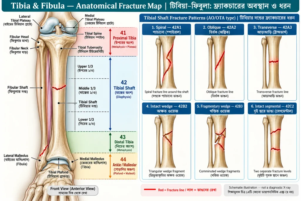
Figure 1 (চিত্র ১): Tibia–Fibula Anatomy with labelled fracture levels and tibial shaft patterns — টিবিয়া–ফিবুলার অ্যানাটমি, Upper/Middle/Lower Third ও ভাঙনের ধরন। ছবির লাল রেখা Fracture বোঝায়; এটি বাস্তব রোগীর X-ray নয়।
| Anatomical level (অবস্থান) | Label / classification (লেবেল) | Fracture examples (উদাহরণ) |
|---|---|---|
| Proximal Tibia — ওপরের টিবিয়া | AO/OTA 41 | Tibial plateau, tibial spine, tibial tuberosity, proximal metaphysis — প্লাটো, স্পাইন, টিউবেরোসিটি ও মেটাফাইসিস |
| Tibial Shaft — মাঝের লম্বা অংশ | AO/OTA 42 | Simple, wedge, segmental/multifragmentary — সরল, ওয়েজ, সেগমেন্টাল |
| Distal Tibia — নিচের টিবিয়া | AO/OTA 43 | Distal extra-articular, partial articular, complete articular / pilon — গোড়ালির ওপরের ও জয়েন্ট-সম্পৃক্ত ফ্র্যাকচার |
| Malleolar / Ankle region — গোড়ালির অঞ্চল | AO/OTA 44 | Malleolar fractures — মেডিয়াল/ল্যাটেরাল/পোস্টেরিয়র ম্যালিওলাসের ফ্র্যাকচার |
| Proximal Fibula — ফিবুলার মাথা ও ঘাড় | Head / neck | Fibular head or neck fracture — ফিবুলার ওপরের অংশ |
| Fibular Shaft — ফিবুলার দণ্ড | Proximal / middle / distal third | Isolated বা associated shaft fracture — একা বা টিবিয়ার সঙ্গে |
| Distal Fibula — গোড়ালির বাইরের হাড় | Lateral malleolus; Weber A/B/C | Syndesmosis-এর নিচে, সমান স্তরে বা ওপরে ফ্র্যাকচার |
Level terminology (অবস্থানের ভাষা): Upper third (ওপরের ১/৩), Middle third (মাঝের ১/৩), Lower/Distal third (নিচের ১/৩)। এগুলো Shaft-এর ভেতরে Fracture Location বর্ণনায় ব্যবহৃত হয়; AO/OTA 41, 42, 43-এর Anatomical Segment Boundary ঠিক Upper/Middle/Lower Third-এর সমার্থক নয়।
Surface landmarks (হাড়ের অংশ): Lateral/Medial Tibial Plateau — হাঁটুর বাইরের/ভেতরের টিবিয়াল পৃষ্ঠ; Tibial Spine — টিবিয়ার মধ্যবর্তী উঁচু অংশ; Tibial Tuberosity — Patellar Tendon-এর সংযুক্তিস্থল; Fibular Head/Neck — ফিবুলার মাথা/ঘাড়; Tibial Shaft — টিবিয়ার দণ্ড; Tibial Plafond — গোড়ালির ওপরের Joint Surface; Medial Malleolus — গোড়ালির ভেতরের হাড়; Lateral Malleolus — গোড়ালির বাইরের হাড়।
3. Imaging Rules and Prescription Format — এক্স-রে নির্বাচন ও প্রেসক্রিপশন লেখার নিয়ম
Standard order format (সাধারণ রীতি): Investigation + Side + Anatomical Region + Views ± Clinical Indication — পরীক্ষার নাম + ডান/বাম + নির্দিষ্ট অংশ + ভিউ + প্রয়োজনে কেন পরীক্ষা।
AP = Anteroposterior View — সামনে-পেছনের অভিমুখে X-ray; Lateral View — পাশ থেকে X-ray; Mortise View — গোড়ালির Joint Mortise দেখার বিশেষ View; Oblique View — তির্যক ভিউ।
| Suspected injury (সন্দেহজনক আঘাত) | Prescription-ready imaging order (প্রেসক্রিপশনের নমুনা) | Why (কেন) |
|---|---|---|
| Tibial plateau/spine/tuberosity — হাঁটুর কাছের টিবিয়া | X-ray Right Knee — AP and Lateral views | Plateau, avulsion, alignment — জয়েন্টের ওপরের অংশ ও বিচ্ছিন্ন অংশ |
| Tibial or fibular shaft — মাঝের অংশ | X-ray Right Leg (Tibia and Fibula) — AP and Lateral views, including knee and ankle joints | Shaft, adjacent joints — দণ্ড ও দুই পাশের জয়েন্ট |
| Proximal fibula — ফিবুলার ওপরের অংশ | X-ray Right Knee — AP and Lateral views | Head/neck fracture — মাথা/ঘাড়ের ফ্র্যাকচার |
| Distal tibia/ankle — গোড়ালির অঞ্চল | X-ray Right Ankle — AP, Lateral and Mortise views | Articular/malleolar injury — জয়েন্ট ও ম্যালিওলাস |
| Pilon / complex plateau — জটিল জয়েন্ট ফ্র্যাকচার | CT Right Ankle / CT Right Knee without IV contrast, if indicated | Articular mapping — ভাঙা Joint Surface-এর গঠন |
| Stress fracture, X-ray negative — স্ট্রেস ফ্র্যাকচার, এক্স-রে স্বাভাবিক | MRI Right Tibia/Fibula (area of concern) without IV contrast, if indicated | Occult bone stress injury — লুকানো হাড়ের ক্ষতি |
| Suspected Maisonneuve injury — মেইসোনিউভ | X-ray Right Ankle — AP, Lateral and Mortise views; X-ray Right Leg (Tibia/Fibula) — AP and Lateral views | Ankle + proximal fibula — গোড়ালি ও ফিবুলার ওপরের অংশ |
Right/Left (ডান/বাম): নমুনায় Right দেওয়া আছে; রোগীর আঘাত Left হলে Left লিখতে হবে। Clinical caution (সতর্কতা): কেবল X-ray দিয়ে Open/Closed Status, Ligament Stability বা Neurovascular Injury নিশ্চিত হয় না; Clinical Examination জরুরি। Acute Ankle Injury-তে উপযুক্ত রোগীর ক্ষেত্রে Ottawa Ankle Rules ব্যবহার করা যায়।
4. Tibial Plateau Fracture: Schatzker I–VI — হাঁটুর টিবিয়াল প্লাটোর ছয় ধরন
Clinical definition (সংজ্ঞা): An intra-articular fracture of the proximal tibial articular surface. বাংলা: হাঁটুর Joint Surface গঠনকারী Tibia-এর ওপরের অংশে ফ্র্যাকচার।
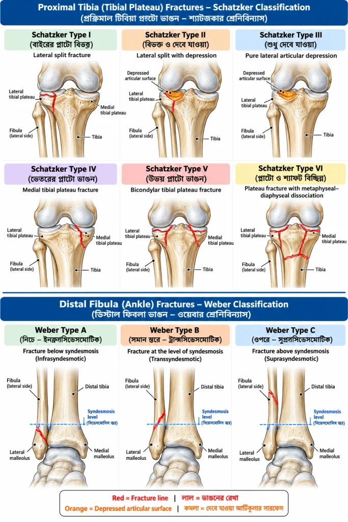
Figure 2 (চিত্র ২): Upper panels: Schatzker Type I–VI (টিবিয়াল প্লাটোর ছয় ধরন); lower panels: Weber A–C (গোড়ালির ফিবুলার তিন ধরন)। Red = fracture line — লাল = ভাঙনের রেখা; Orange = articular depression — কমলা = দেবে যাওয়া জয়েন্টের পৃষ্ঠ। চিত্র শিক্ষামূলক।
Common investigations (প্রাথমিক পরীক্ষা): X-ray Right Knee — AP and Lateral views। Selected additional study (প্রয়োজনে): CT Right Knee without IV contrast — Articular Depression ও Fracture Mapping বোঝার জন্য। Meniscal/Ligament Injury সন্দেহ হলে নির্বাচিত ক্ষেত্রে MRI।
Schatzker Type I — Lateral Split Fracture (বাইরের প্লাটো বিভক্ত)
Definition: A split fracture of the lateral tibial plateau without major depression. বাংলা: টিবিয়াল প্লাটোর বাইরের অংশে বিভক্ত রেখা তৈরি হয়েছে, উল্লেখযোগ্য Depression নেই। X-ray: Lateral split line — বাইরের পৃষ্ঠে বিভক্ত রেখা। Conservative: Undisplaced/minimally displaced ও clinically stable knee হলে Hinged Brace, Restricted Weight Bearing ও Serial Imaging। OT: Displacement, Articular Incongruity অথবা Instability থাকলে Lag Screw/Plate Fixation (ORIF)।
Schatzker Type II — Lateral Split-Depression (বিভক্ত ও দেবে যাওয়া)
Definition: Lateral plateau split associated with depressed articular surface. বাংলা: বাইরের অংশ বিভক্ত, পাশাপাশি জয়েন্টের পৃষ্ঠ নিচে বসে গেছে। X-ray/CT: Split এবং Depression; CT-তে আকার স্পষ্ট। Conservative: Stable, minimally displaced/depressed নির্বাচিত ক্ষেত্রে। OT: Significant Depression/Incongruity/Instability হলে Articular Elevation, Bone-defect Support ও ORIF with Plate/Screws।
Schatzker Type III — Pure Lateral Depression (শুধু বাইরের পৃষ্ঠ দেবে যাওয়া)
Definition: Depression of lateral tibial articular surface without a major split. বাংলা: বড় বিভক্ত অংশ ছাড়া জয়েন্টের বাইরের পৃষ্ঠ নিচে দেবে গেছে। X-ray/CT: Subchondral Depression — পৃষ্ঠের নিচের হাড় বসে যাওয়া। Conservative: Minimal Depression, stable alignment। OT: Significant Joint Depression/Incongruity হলে Elevation and Internal Fixation।
Schatzker Type IV — Medial Plateau Fracture (ভেতরের প্লাটো ভাঙা)
Definition: Fracture involving the medial tibial plateau. বাংলা: টিবিয়াল প্লাটোর ভেতরের অংশ ভেঙে যাওয়া। X-ray/CT: Medial Articular Fracture; উচ্চ-শক্তির আঘাতে Associated Vascular/Ligament Injury খুঁজতে হবে। Conservative: Selected nondisplaced stable fracture। OT: Displaced/unstable injury-তে Medial/Posteromedial Buttress Plate Fixation।
Schatzker Type V — Bicondylar Plateau (দুই দিকের প্লাটো ভাঙা)
Definition: Fracture involving medial and lateral tibial plateaus. বাংলা: প্লাটোর ভেতরের ও বাইরের দুই দিকই ভেঙেছে। X-ray/CT: Bicondylar Fragments — দুই কন্ডাইলে ভাঙা অংশ। Conservative: বিরল, স্থিতিশীল ও Minimally Displaced নির্বাচিত ক্ষেত্রে। OT: Displaced/Unstable হলে ORIF, কখনও Dual-Column Fixation; Soft Tissue Swelling বেশি হলে Staged Surgery।
Schatzker Type VI — Metaphyseal–Diaphyseal Dissociation (প্লাটো ও শ্যাফট বিচ্ছিন্ন)
Definition: A plateau fracture with dissociation between the articular segment and tibial diaphysis. বাংলা: হাঁটুর Joint-Bearing অংশ Shaft থেকে ভেঙে বিচ্ছিন্ন। X-ray: Plateau Fracture with Metaphyseal/Shaft Discontinuity। Conservative: বিরল Selected Stable Case। OT: বেশিরভাগ Displaced/Unstable Injury-তে Temporary Spanning External Fixation ও পরে Definitive Plate Fixation/Selected Ring Fixation।
Clinical point (চিকিৎসাগত কথা): Schatzker Grade একা Surgery নির্ধারণ করে না। Articular Congruity, Stability, Soft Tissue, Vascular Status ও রোগীর অবস্থা বিবেচনা জরুরি।
5. Other Proximal Tibia and Fibula Injuries — হাঁটুর কাছের অন্যান্য ফ্র্যাকচার
Tibial Spine / Intercondylar Eminence Avulsion (টিবিয়াল স্পাইনের অ্যাভালশন)
Definition: Avulsion of the tibial intercondylar eminence, often at the ACL attachment. বাংলা: ACL যেখানে যুক্ত, সেই টিবিয়াল উঁচু অংশ টানে বিচ্ছিন্ন হওয়া। X-ray order: X-ray Right Knee — AP and Lateral views; নির্বাচিত ক্ষেত্রে MRI/CT। Finding: Eminence Fragment — আলাদা ছোট হাড়ের অংশ। Conservative: Nondisplaced/stably reduced হলে immobilization। OT: Displaced, irreducible, entrapped fragment বা persistent instability-তে arthroscopic/open fixation।
Tibial Tuberosity Avulsion (টিবিয়াল টিউবেরোসিটির অ্যাভালশন)
Definition: Avulsion at the patellar tendon insertion on the tibial tuberosity. বাংলা: প্যাটেলার টেনডনের টানে সামনের উঁচু টিবিয়াল অংশ ভাঙা। X-ray order: X-ray Right Knee — AP and Lateral views (Lateral বিশেষ গুরুত্বপূর্ণ)। Finding: Displaced Tuberosity Fragment — সামনের হাড়ের টুকরা সরে যাওয়া। Conservative: Nondisplaced এবং extensor mechanism intact হলে extension immobilization। OT: Displaced অথবা extensor disruption হলে ORIF/Screw Fixation।
Proximal Tibial Metaphyseal Fracture (টিবিয়ার ওপরের মেটাফাইসিসের ফ্র্যাকচার)
Definition: Fracture of proximal tibial metaphysis, below the articular plateau. বাংলা: প্লাটোর ঠিক নিচের প্রসারিত অংশ ভাঙা। X-ray order: X-ray Right Knee — AP and Lateral views, প্রয়োজনে Tibia/Fibula Views। Conservative: Stable ও Acceptably Aligned হলে Cast/Brace। OT: Unstable, Displaced অথবা reduction না টিকলে Plate/Nail/External Fixation, নির্বাচিত ক্ষেত্রে।
Fibular Head or Neck Fracture (ফিবুলার মাথা বা ঘাড়ের ফ্র্যাকচার)
Definition: Fracture at the proximal fibular head or neck. বাংলা: ফিবুলার ওপরের Head বা Neck ভাঙা। X-ray order: X-ray Right Knee — AP and Lateral views; Ankle Injury থাকলে পূর্ণ Lower-Leg/Ankle Imaging। Conservative: Isolated stable fracture। OT: Associated Knee Ligament Injury/Syndesmotic Instability বা selected displaced avulsion হলে সংশ্লিষ্ট Structure-এর Surgery। Warning: Common Fibular Nerve পরীক্ষা করতে হবে; Foot Drop হতে পারে।
6. Tibial Shaft Fracture: AO/OTA 42 — টিবিয়ার দণ্ডের ফ্র্যাকচার
Definition: Fracture involving the tibial diaphysis. বাংলা: টিবিয়ার মাঝের লম্বা দণ্ড ভেঙে যাওয়া। Prescription: X-ray Right Leg (Tibia and Fibula) — AP and Lateral views, including knee and ankle joints। CT: Isolated shaft fracture-এ routine নয়; Joint Extension সন্দেহে।
| AO/OTA type — শ্রেণি | Clinical definition — ইংরেজি ও বাংলা | Typical radiographic finding — X-ray-তে | Conservative — অপারেশন ছাড়া | OT / operation — কখন ও কী |
|---|---|---|---|---|
| 42-A1 Simple spiral — প্যাঁচানো | Helical fracture line; বাংলা: দণ্ড ঘিরে প্যাঁচানো ভাঙন | Spiral line — সর্পিল রেখা | Stable/acceptable alignment: cast/functional brace — স্থিতিশীল হলে প্লাস্টার/ব্রেস | Unstable/displaced/rotation: IM interlocking nail; selected plate — অস্থিতিশীল হলে নেইল |
| 42-A2 Simple oblique — তির্যক | Single oblique line; বাংলা: একটিমাত্র তির্যক ভাঙন | Oblique line — তির্যক রেখা | Stable/reduced: cast — স্থিতিশীল হলে প্লাস্টার | Unstable/shortening: IM nail or plate — সরে গেলে নেইল/প্লেট |
| 42-A3 Simple transverse — আড়াআড়ি | Line approximately across shaft; বাংলা: প্রায় আড়াআড়িভাবে ভাঙা | Transverse line — আড়াআড়ি রেখা | Stable: cast — স্থিতিশীল হলে প্লাস্টার | Displaced/unacceptable angulation: IM nail — বিকৃতি হলে নেইল |
| 42-B2 Intact wedge — অক্ষত ওয়েজ | Single intact wedge between main fragments; বাংলা: মাঝখানে অক্ষত ত্রিভুজাকার অংশ | Wedge/butterfly fragment — ওয়েজ টুকরা | Selected stable case — নির্বাচিত স্থিতিশীল আঘাত | Unstable: IM nail/bridge plate — নেইল/ব্রিজ প্লেট |
| 42-B3 Fragmentary wedge — খণ্ডিত ওয়েজ | Wedge component is fragmented; বাংলা: মাঝের ওয়েজ একাধিক টুকরা | Fragmented wedge — খণ্ডিত ওয়েজ | Rare selected stable case — বিরল ক্ষেত্রে | Displaced/unstable: IM nail/bridge plate |
| 42-C2 Intact segmental — অক্ষত সেগমেন্ট | Two fracture levels with intact middle segment; বাংলা: দুই স্তরে ভেঙে মাঝখানে আলাদা অক্ষত অংশ | Two distinct breaks — দুই স্থানে ভাঙা | Highly selected cases — খুব নির্বাচিত | Most unstable cases: IM nail/appropriate fixation |
| 42-C3 Fragmentary segmental — খণ্ডিত সেগমেন্ট | Intermediate segment fragmented; বাংলা: মাঝের সেগমেন্টও টুকরা | Multiple segmental fragments — বহু টুকরা | Generally unsuitable if unstable — অস্থিতিশীল হলে সাধারণত নয় | IM nail, bridge plate, ring/external fixation according to soft tissue |
Treatment caveat (চিকিৎসা সতর্কতা): Adult Tibial Shaft-এর বহু Operative Case-এ IM Nailing গুরুত্বপূর্ণ, কিন্তু Fracture Level, Soft Tissue ও Joint Extension অনুযায়ী Plate বা External Fixator বেশি উপযুক্ত হতে পারে। Open Fracture ও Compartment Syndrome আলাদা Emergency Priority।
7. Distal Tibia, Pilon and AO/OTA 43 — টিবিয়ার নিচের অংশ ও পাইলন
Distal Tibial Metaphyseal Fracture (নিচের মেটাফাইসিস ভাঙা)
Definition: Fracture of distal tibial metaphysis, with or without articular extension. বাংলা: গোড়ালির কাছের টিবিয়ার বিস্তৃত অংশ ভাঙা, কখনও Joint Surface পর্যন্ত পৌঁছায়। X-ray order: X-ray Right Leg (Tibia and Fibula) — AP and Lateral views; Joint involvement থাকলে X-ray Right Ankle — AP, Lateral and Mortise views। Conservative: Stable, extra-articular, acceptably aligned। OT: Unstable/Displaced হলে Selected IM Nail, Plate or External Fixation।
Tibial Pilon / Plafond Fracture (টিবিয়াল পাইলন—গোড়ালির ওপরের জয়েন্ট পৃষ্ঠ ভাঙা)
Definition: An intra-articular fracture of the weight-bearing distal tibial plafond, often from axial loading. বাংলা: গোড়ালির ওপরের ওজনবাহী টিবিয়াল জয়েন্টের পৃষ্ঠ ভাঙা। X-ray order: X-ray Right Ankle — AP, Lateral and Mortise views; CT: CT Right Ankle without IV contrast for planning। X-ray/CT: Articular Step, Depression, Comminution — জয়েন্টে উঁচু-নিচু, বসে যাওয়া ও টুকরা। Conservative: Nondisplaced stable selected cases। OT: Displaced Articular Injury/Instability; severe swelling হলে Span–Scan–Plan (temporary external fixator → CT → definitive ORIF when soft tissue allows)।
| AO/OTA 43 type | Definition — বাংলা অর্থ | Typical management — চিকিৎসা |
|---|---|---|
| 43-A Extra-articular — জয়েন্টের বাইরে | Distal segment fracture without joint surface involvement — জয়েন্টের পৃষ্ঠ অক্ষত | Stable: cast; unstable: nail/plate — স্থিতিশীল হলে প্লাস্টার, না হলে ফিক্সেশন |
| 43-B Partial articular — আংশিক জয়েন্ট-সম্পৃক্ত | Part of articular surface remains attached to shaft — পৃষ্ঠের কিছু অংশ Shaft-এর সঙ্গে যুক্ত | Minimal displacement/stable: selected conservative; incongruity: ORIF |
| 43-C Complete articular — সম্পূর্ণ জয়েন্ট-সম্পৃক্ত | Articular segment separated from shaft — জয়েন্ট-বহনকারী অংশ Shaft থেকে বিচ্ছিন্ন | Displaced/unstable: usually surgical reconstruction/fixation — সাধারণত অস্ত্রোপচার |
Classification clarification (শ্রেণিবিন্যাস): AO/OTA 43 distal tibial end-এর Articular/Extra-articular Fracture Group; 44 malleolar ankle region-এর Fracture Group। Anatomical Terms ও Code একে অপরের বিকল্প নয়।
8. Malleolar and Distal Fibular Fractures: Weber A–C — গোড়ালির ফিবুলা ও ম্যালিওলাস
Medial Malleolus Fracture (গোড়ালির ভেতরের হাড় ভাঙা)
Definition: Fracture of the distal tibial medial malleolus. বাংলা: টিবিয়ার নিচের ভেতরের উঁচু হাড় ভাঙা। X-ray: X-ray Right Ankle — AP, Lateral and Mortise views। Conservative: Stable, nondisplaced হলে Cast/Boot। OT: Displacement/unstable ankle-এ Lag Screws, Tension Band বা Buttress Plate (pattern অনুযায়ী)।
Posterior Malleolus Fracture (গোড়ালির পেছনের হাড় ভাঙা)
Definition: Fracture of the posterior articular margin of distal tibia. বাংলা: গোড়ালির Joint Surface-এর পেছনের অংশ ভাঙা। X-ray: Ankle AP/Lateral/Mortise; CT: Fragment, Impaction, Syndesmosis বোঝার জন্য প্রায়ই উপকারী। Conservative: Small, nondisplaced, congruent stable ankle। OT: Displaced/unstable/incongruent joint-এ Screw বা Posterior Buttress Plate। শুধু Fragment Size দেখে নয়—Stability এবং CT Morphology-ও গুরুত্বপূর্ণ।
Lateral Malleolus / Distal Fibula Fracture (গোড়ালির বাইরের ফিবুলা ভাঙা)
Definition: Fracture of the distal fibula forming the lateral ankle malleolus. বাংলা: গোড়ালির বাইরের ফিবুলার শেষ প্রান্ত ভাঙা। X-ray: X-ray Right Ankle — AP, Lateral and Mortise views। Conservative/OT: Mortise Stability এবং Associated Medial/Syndesmotic Injury দেখে।
| Weber label (লেবেল) | Clinical definition — ইংরেজি + বাংলা | Conservative — অপারেশন ছাড়া | OT / operative — অস্ত্রোপচার |
|---|---|---|---|
| Weber A — Infrasyndesmotic | Fracture below syndesmosis — Syndesmosis-এর নিচে ফিবুলা ভাঙা | Stable isolated ankle: walking boot/functional support, WBAT if permitted — স্থিতিশীল হলে বুট | Unstable/associated injury: selected ORIF — অস্থিতিশীল হলে ফিক্সেশন |
| Weber B — Transsyndesmotic | Fracture at syndesmosis — Syndesmosis-এর সমান স্তরে ফিবুলা ভাঙা | Stable mortise: boot/cast; supervised weight-bearing radiograph sometimes helpful — স্থিতিশীল হলে বুট/প্লাস্টার | Medial instability/talar shift: fibular ORIF ± syndesmotic fixation — সরে গেলে ফিক্সেশন |
| Weber C — Suprasyndesmotic | Fracture above syndesmosis — Syndesmosis-এর ওপরে ভাঙা | Rare, proven stable selected case — বিরল স্থিতিশীল ক্ষেত্রে | Syndesmotic instability/mortise incongruity: reduction and syndesmotic stabilization ± fibular fixation |
Weber figure label (ছবির লেবেল): চিত্র ২-এর নিচের অংশে A = below, B = at, C = above দেখানো হয়েছে। Weber Type একা Ankle Stability নিশ্চিত করে না।
Maisonneuve Injury (মেইসোনিউভ আঘাত)
Definition: Proximal fibular fracture with distal tibiofibular syndesmotic disruption and medial-sided ankle injury. বাংলা: ফিবুলার ওপরের অংশ ভাঙার সঙ্গে গোড়ালির Syndesmosis ও ভেতরের Stabilizing Structure ক্ষতিগ্রস্ত হওয়া। Orders: X-ray Right Ankle — AP, Lateral and Mortise views + X-ray Right Leg (Tibia/Fibula) — AP and Lateral views। Conservative: বিরল প্রমাণিত Stable Case। OT: Unstable syndesmosis-এ ankle reduction/syndesmotic fixation; proximal fibula-তে সবসময় Plate লাগে না।
Isolated Fibular Shaft Fracture (শুধু ফিবুলার দণ্ড ভাঙা)
Definition: Fibular diaphyseal fracture without unstable associated injury. বাংলা: অন্য অস্থিতিশীল আঘাত ছাড়া শুধু Fibula Shaft ভাঙা। X-ray: X-ray Right Leg (Tibia and Fibula) — AP and Lateral views। Conservative: বেশিরভাগ Stable Case-এ Boot/Support ও Pain-guided Rehabilitation। OT: Isolated Stable Case-এ সাধারণত নয়; Associated Tibial/Ankle Injury থাকলে আলাদা সিদ্ধান্ত।
9. Fracture Morphology: Other Patterns — অন্যান্য ভাঙনের আকৃতি
| Type — ধরন | Clinical definition — সংজ্ঞা ও বাংলা | Imaging appearance — এক্স-রেতে | Treatment principle — চিকিৎসা |
|---|---|---|---|
| Transverse — আড়াআড়ি | Break roughly perpendicular to long axis — লম্বা অক্ষের প্রায় সমকোণে ভাঙা | Crosswise line — আড়াআড়ি রেখা | Stable: cast; unstable: fixation |
| Oblique — তির্যক | Angled fracture line — তির্যক রেখায় ভাঙা | Slant line — কাত রেখা | Alignment/rotation অনুযায়ী Cast বা OT |
| Spiral — সর্পিল | Helical fracture line — হাড় ঘিরে প্যাঁচানো রেখা | Helical line — সর্পিল রেখা | Stable Cast; unstable IM Nail/plate |
| Comminuted — বহুখণ্ডিত | At least three bone fragments — অন্তত তিন টুকরা | Multiple fragments — বহু টুকরা | Stability/soft tissue অনুযায়ী |
| Segmental — সেগমেন্টাল | Two separate levels leaving intermediate segment — দুই স্তরে ভেঙে মাঝখানে অংশ | Two levels — দুই ভাঙা স্থান | Displaced/unstable হলে প্রায়ই OT |
| Avulsion — অ্যাভালশন | Tendon/ligament pulls bone fragment — টেনডন/লিগামেন্টের টানে অংশ বিচ্ছিন্ন | Small detached fragment — আলাদা টুকরা | Stable Cast/Brace; displaced functional injury OT |
| Impacted — চাপা পড়া | Fragment driven into another — এক অংশ অন্যটির মধ্যে ঢুকে যায় | Impaction/shortening — ঘনত্ব/খাটো | Congruity/alignment দেখে |
| Greenstick — আংশিক ভাঙন | One cortex breaks, other bends — একপাশ ভাঙে, অন্যপাশ বাঁকে | Incomplete cortical break — অসম্পূর্ণ ভাঙন | শিশুতে সাধারণত Cast/Reduction |
| Buckle/Torus — কর্টেক্স বেঁকে যাওয়া | Cortical buckling from compression — চাপের কারণে Cortex ফুলে/বেঁকে যায় | Cortical bulge — কর্টেক্সের ফোলা | সাধারণত Support/Immobilization |
| Stress fracture — পুনরাবৃত্ত চাপজনিত | Bone injury due to repeated loading or weak bone — বারবার চাপ বা হাড় দুর্বলতায় ভাঙা | Early X-ray may be normal — প্রথমে স্বাভাবিক হতে পারে | Activity modification; MRI if X-ray negative; high-risk site review |
| Pathological — রোগজনিত | Break through disease-weakened bone — রোগে দুর্বল হাড় ভাঙা | Abnormal bone +/- fracture — অস্বাভাবিক হাড় | Etiology work-up আগে; Tumor সন্দেহে specialist plan |
Pattern ≠ treatment decision (আকৃতি ≠ একমাত্র অপারেশনের কারণ): Morphology-এর সঙ্গে Displacement, Open Wound, Joint Congruity, Stability, Age ও Neurovascular Status দেখতে হবে।
10. Open/Closed Fracture and Gustilo Grade — খোলা/বন্ধ ফ্র্যাকচার
Closed fracture — বন্ধ ফ্র্যাকচার: Fracture without communication with outside environment. বাংলা: Fracture Site-এর সঙ্গে বাইরের পরিবেশের যোগাযোগ নেই।
Open fracture — খোলা ফ্র্যাকচার: Fracture communicating with outside via skin wound. বাংলা: ত্বকের ক্ষত দিয়ে ভাঙা অংশ বাইরের পরিবেশের সঙ্গে যোগাযোগ করে; হাড় বাইরে দেখা জরুরি নয়।
| Gustilo type (গ্রেড) | Clinical meaning — বাংলা অর্থ |
|---|---|
| I | Small, relatively clean wound, limited soft-tissue injury — ছোট অপেক্ষাকৃত পরিষ্কার ক্ষত |
| II | Larger wound without extensive soft-tissue destruction — বড় ক্ষত কিন্তু ব্যাপক টিস্যু ক্ষতি নেই |
| IIIA | Severe injury yet adequate bone coverage — গুরুতর আঘাত হলেও হাড় ঢাকার টিস্যু আছে |
| IIIB | Extensive soft-tissue loss, often flap required — ব্যাপক টিস্যু ক্ষতি, Flap লাগতে পারে |
| IIIC | Arterial injury requiring repair — ধমনি ক্ষতিগ্রস্ত, Repair দরকার |
Important (গুরুত্বপূর্ণ): Gustilo Grade কেবল ক্ষতের সেন্টিমিটার মেপে নির্ধারণ করা যায় না; Tissue Damage ও operative assessment গুরুত্বপূর্ণ। Initial management (প্রাথমিক ব্যবস্থা): Trauma ABC, urgent neurovascular evaluation, sterile dressing, splint, early IV antibiotics, tetanus assessment এবং urgent orthopedic/orthoplastic referral। Operation (অস্ত্রোপচার): Surgical wound excision/debridement, stabilization, vascular repair/soft-tissue coverage as needed। Emergency condition—শুধু Cast যথেষ্ট নয়।
11. Displacement, Alignment and Joint Involvement — ভাঙা অংশের অবস্থান
| Term (শব্দ) | Clinical meaning — বাংলা অর্থ | Clinical relevance — গুরুত্ব |
|---|---|---|
| Nondisplaced — অস্থানচ্যুত | Bone fragments remain aligned — ভাঙলেও অংশ স্বাভাবিক রেখায় | Stable হলে Conservative সম্ভব |
| Displaced — স্থানচ্যুত | Fragments shift/translate — অংশ সরে যায় | Reduction ও stability assessment |
| Angulated — কোণ তৈরি | Abnormal angle at fracture — অস্বাভাবিক কোণ | Unacceptable হলে Correction |
| Rotated — ঘূর্ণিত | One fragment rotated — অংশ ঘুরে গেছে | Malrotation correction গুরুত্বপূর্ণ |
| Shortened — ছোট | Overlap/loss of length — হাড়ের দৈর্ঘ্য কম | উল্লেখযোগ্য হলে Reduction |
| Distracted — ফাঁক তৈরি | Excessive gap between pieces — অংশের মধ্যে ফাঁক | Healing/stability প্রভাবিত |
| Intra-articular — জয়েন্ট-সম্পৃক্ত | Fracture reaches joint surface — জয়েন্টের পৃষ্ঠ পর্যন্ত | Congruity গুরুত্বপূর্ণ |
| Extra-articular — জয়েন্টের বাইরে | Joint surface not involved — জয়েন্টের পৃষ্ঠ অক্ষত | Alignment/Stability মূল বিষয় |
Neurovascular status (স্নায়ু ও রক্তনালির অবস্থা) এবং Open/Closed status (ক্ষত খোলা/বন্ধ) অবশ্যই Clinical Examination-এ লিখতে হবে; X-ray Alone যথেষ্ট নয়।
12. Pediatric and Growth-Plate Fractures — শিশুদের ফ্র্যাকচার
Toddler’s fracture — টডলার ফ্র্যাকচার: Usually a nondisplaced spiral/oblique distal tibial fracture in an ambulatory toddler — হাঁটতে শেখা ছোট শিশুর নিচের টিবিয়ায় সূক্ষ্ম ভাঙন। Order: X-ray Right Leg (Tibia and Fibula) — AP and Lateral views ± Oblique; প্রথম X-ray স্বাভাবিক হতে পারে। Treatment: সাধারণত Conservative Immobilization/Support, Follow-up।
| Salter–Harris type | Clinical definition — বাংলা অর্থ | Management principle — চিকিৎসা |
|---|---|---|
| I | Through physis — কেবল Growth Plate দিয়ে | Nondisplaced: cast; displaced: reduction |
| II | Physis + metaphysis — গ্রোথ প্লেট ও মেটাফাইসিস | Stable Cast; displaced Reduction |
| III | Physis + epiphysis, intra-articular — গ্রোথ প্লেট ও জয়েন্টের দিক | Articular congruity; displaced often fixation |
| IV | Metaphysis + physis + epiphysis — তিন অংশ জড়িত | Anatomical Reduction গুরুত্বপূর্ণ |
| V | Crush injury of growth plate — গ্রোথ প্লেট চাপে ক্ষতিগ্রস্ত | Specialist follow-up; growth arrest risk |
Distal pediatric ankle X-ray: X-ray Right Ankle — AP, Lateral and Mortise views। Tillaux fracture — টিলো: Transitional Salter–Harris III anterolateral distal tibial epiphysis (কৈশোরে টিবিয়ার নিচের বাইরের এপিফাইসিস ভাঙা)। Triplane fracture — ট্রাইপ্লেন: Transitional fracture through three planes (তিনটি প্লেনে ভাঙা)। CT: selected cases to measure articular displacement। Management: <2 mm and stable may be casted; >2 mm displacement typically warrants orthopedic operative assessment; physeal follow-up জরুরি।
13. Causes, Symptoms and Emergency Examination — কারণ, লক্ষণ ও জরুরি পরীক্ষা
Mechanisms (কারণ): Road traffic collision — সড়ক দুর্ঘটনা; Fall from height — উঁচু থেকে পড়া; Direct blow — সরাসরি আঘাত; Twisting injury — প্যাঁচানো আঘাত; Sports injury — খেলাধুলায় আঘাত; Repetitive loading — বারবার চাপ; Osteoporosis/pathological bone — দুর্বল বা রোগাক্রান্ত হাড়।
Symptoms (লক্ষণ): Pain — ব্যথা; Swelling — ফোলা; Tenderness — চাপ দিলে ব্যথা; Bruising — কালশিটে; Deformity — বিকৃতি; Inability to bear weight — ভর দিতে না পারা; Reduced movement — নড়াচড়া কম; Numbness/Foot drop — অনুভূতি কমে যাওয়া/পায়ের পাতা তুলতে না পারা।
Clinical examination (পরীক্ষা): Skin integrity/open wound — ক্ষত দেখা; Compartments — পেশির চাপের লক্ষণ; Dorsalis pedis & posterior tibial pulses — পায়ের ধমনির স্পন্দন; Capillary refill — রক্তফেরার সময়; Motor/sensation — নড়াচড়া/অনুভূতি; adjacent Knee and Ankle — হাঁটু ও গোড়ালি পরীক্ষা। Pre- and post-reduction neurovascular findings লিখতে হবে।
Acute compartment syndrome — তীব্র কম্পার্টমেন্ট সিনড্রোম: Increased pressure in muscle compartment compromises perfusion — পেশির আবদ্ধ অংশে চাপ বেড়ে রক্ত সরবরাহ ব্যাহত। Warning: Pain out of proportion — অস্বাভাবিক তীব্র ব্যথা; Pain on passive stretch — পেশি টানলে ব্যথা; tense swelling — শক্ত ফোলা; progressive paresthesia/weakness — ঝিনঝিনি বা দুর্বলতা বাড়া। Emergency: Urgent surgical assessment and fasciotomy when indicated — জরুরি অপারেশন মূল্যায়ন; প্রয়োজনে ফ্যাসিওটমি। Pulse থাকলেও এটি বাদ যায় না।
14. Prescription-Ready Clinical Examples — বাস্তব প্রেসক্রিপশনের নমুনা
Example A — Suspected Tibial Shaft Fracture (টিবিয়ার দণ্ড ভাঙার সন্দেহ)
Provisional Dx: ? Fracture shaft of right tibia following trauma — আঘাতের পর ডান টিবিয়ার দণ্ডে ফ্র্যাকচারের সন্দেহ।
Investigation: X-ray Right Leg (Tibia and Fibula) — AP and Lateral views, including knee and ankle joints.
Purpose: Identify level, pattern and alignment — অবস্থান, ধরন ও সরে যাওয়ার মাত্রা বোঝা।
Example B — Suspected Tibial Plateau Fracture (টিবিয়াল প্লাটোর সন্দেহ)
Provisional Dx: ? Right tibial plateau fracture।
Investigation: X-ray Right Knee — AP and Lateral views.
Additional: CT Right Knee without IV contrast, if fracture configuration/articular depression requires clarification.
Example C — Suspected Left Ankle Fracture (বাম গোড়ালি ভাঙার সন্দেহ)
Provisional Dx: ? Left ankle fracture।
Investigation: X-ray Left Ankle — AP, Lateral and Mortise views.
Purpose: Assess malleoli and ankle mortise — গোড়ালির হাড় ও জয়েন্টের সমতা।
Example D — Maisonneuve Pattern (মেইসোনিউভের সন্দেহ)
Provisional Dx: ? Right Maisonneuve injury / syndesmotic disruption।
Investigations: X-ray Right Ankle — AP, Lateral and Mortise views plus X-ray Right Leg (Tibia and Fibula) — AP and Lateral views (include proximal fibula).
Example E — Postoperative Union Assessment (অপারেশনের পর হাড় জোড়া লাগার মূল্যায়ন)
Diagnosis: Status post IM interlocking nailing, right tibial shaft fracture.
Investigation: X-ray Right Leg (Tibia and Fibula) — AP and Lateral views.
Indication: Follow-up: fracture alignment, implant position and progression of union. — অক্ষ, ইমপ্লান্ট ও জোড়া লাগা মূল্যায়ন।
Example F — Suspected Stress Fracture (স্ট্রেস ফ্র্যাকচার সন্দেহ)
Investigation: X-ray Right Leg (Tibia and Fibula) — AP and Lateral views। X-ray negative/indeterminate কিন্তু সন্দেহ থাকলে MRI Right Tibia/Fibula (site of pain) without IV contrast।
Prescription wording principles (লেখার নীতি): Right/Left ভুল না করা; Fracture Pattern বা Schatzker/Weber Grade Imaging-এ নিশ্চিত না হলে ?/Suspected লিখতে হবে; Open/Closed Status Clinical Examination অনুযায়ী; CT/MRI সব রোগীতে একসঙ্গে Routine নয়।
15. Specific Treatment: Conservative vs OT — অপারেশন ছাড়া বনাম অস্ত্রোপচার
Initial management — প্রথম ধাপ: Trauma ABC — জীবনরক্ষাকারী মূল্যায়ন; analgesia — ব্যথা নিয়ন্ত্রণ; sterile dressing if wound — ক্ষতে জীবাণুমুক্ত ড্রেসিং; splint/immobilization — স্থির রাখা; neurovascular/compartment reassessment — রক্তনালি ও পেশির চাপ বারবার দেখা; appropriate imaging — উপযুক্ত X-ray। Deformity থাকলে চিকিৎসকের তত্ত্বাবধানে জরুরি Reduction প্রয়োজন হতে পারে।
| Clinical situation — অবস্থা | Conservative (অপারেশন ছাড়া) | OT / Surgical indication (অস্ত্রোপচার) |
|---|---|---|
| Stable aligned shaft fracture — স্থিতিশীল দণ্ড | Cast / Functional Brace + serial X-ray — প্লাস্টার/ব্রেস | Failure of reduction, rotation, instability — অক্ষ নষ্ট হলে Fixation |
| Tibial plateau minimally displaced/stable — প্লাটো স্থিতিশীল | Hinged brace, restricted loading, guided movement — ব্রেস ও নিয়ন্ত্রিত ভর | Articular incongruity/instability — জয়েন্টের অসামঞ্জস্য হলে ORIF |
| Displaced pilon — স্থানচ্যুত পাইলন | Usually not if unstable — অস্থিতিশীল হলে নয় | Staged Ex-Fix / ORIF according to soft tissue — পর্যায়ক্রমে অপারেশন |
| Isolated stable fibular shaft — ফিবুলা একা স্থিতিশীল | Support/boot, symptom-guided activity — সাপোর্ট | Associated unstable injury — অন্য অস্থিতিশীল আঘাত থাকলে |
| Stable Weber A/B — স্থিতিশীল গোড়ালি | Boot/cast, WBAT if appropriate — বুট/প্লাস্টার | Talar shift or medial instability — ট্যালাস সরে গেলে ORIF |
| Unstable Weber B/C/Maisonneuve — অস্থিতিশীল গোড়ালি | Generally not definitive — সাধারণত নয় | Ankle/syndesmotic stabilization — গোড়ালি স্থির করা |
| Open fracture — খোলা ফ্র্যাকচার | Splint alone insufficient — শুধু প্লাস্টার নয় | Urgent antibiotics, wound excision/debridement, stabilization — জরুরি অস্ত্রোপচার ব্যবস্থাপনা |
| Acute compartment syndrome — কম্পার্টমেন্ট সিনড্রোম | Not definitive — নয় | Emergency fasciotomy — জরুরি ফ্যাসিওটমি |
| Major arterial injury — বড় ধমনির আঘাত | Not definitive — নয় | Urgent vascular assessment/repair — জরুরি রক্তনালি চিকিৎসা |
Procedure definitions (অপারেশনের অর্থ): IM interlocking nailing — ইন্ট্রামেডুলারি নেইলিং: Shaft-এর ভেতর Nail ও Locking Screws দিয়ে স্থির করা। ORIF — ওপেন রিডাকশন অ্যান্ড ইন্টারনাল ফিক্সেশন: খোলা অস্ত্রোপচারে হাড়ের অবস্থান ঠিক করে Plate/Screw দিয়ে স্থির করা। External fixation — এক্সটার্নাল ফিক্সেশন: হাড়ে Pin/Wire দিয়ে শরীরের বাইরে Frame বসানো। Ilizarov ring fixation — ইলিজারভ রিং: Circular Frame দিয়ে জটিল ফ্র্যাকচার/ননইউনিয়ন/বিকৃতি চিকিৎসা। Fasciotomy — ফ্যাসিওটমি: পেশির আবদ্ধ অংশের চাপ কমাতে Surgical Release।
Weight-bearing orders (পায়ে ভর দেওয়া): NWB Non-weight-bearing — ভর না দেওয়া; TTWB Toe-touch — আঙুলের হালকা স্পর্শ; PWB Partial — আংশিক ভর; WBAT Weight-bearing as tolerated — চিকিৎসকের অনুমোদন সাপেক্ষে সহ্যক্ষমতা অনুযায়ী; FWB Full weight-bearing — সম্পূর্ণ ভর। Implant থাকলেই FWB নিরাপদ নয়।
16. Healing, Union, Follow-up and Complications — হাড় জোড়া লাগা ও জটিলতা
Fracture healing phases (জোড়া লাগার ধাপ): Inflammation — প্রদাহ; Soft Callus — নরম নতুন টিস্যু; Hard Callus — শক্ত নতুন হাড়; Remodeling — পরিণত হাড়ে পুনর্গঠন। Callus (ক্যালাস): Fracture Healing-এ তৈরি Reparative Tissue — নতুন জোড়া লাগার টিস্যু। Bridging Callus (ব্রিজিং ক্যালাস): ভাঙা দুই অংশের মধ্যে নতুন হাড়ের সংযোগ।
Clinical union (ক্লিনিক্যাল ইউনিয়ন): Pain/Tenderness কমা এবং উপযুক্ত Functional Stability পাওয়া। Radiographic union (রেডিওগ্রাফিক ইউনিয়ন): Serial X-ray-তে পর্যাপ্ত Bridging/Progressive Healing। Delayed union (বিলম্বিত ইউনিয়ন): স্বাভাবিকের তুলনায় ধীর Healing। Nonunion (ননইউনিয়ন): হাড় জোড়া লাগার অগ্রগতি ব্যর্থ। Malunion (ম্যালইউনিয়ন): ভুল Alignment-এ জোড়া লাগা।
Follow-up order (ফলোআপ পরীক্ষা): X-ray Right Leg (Tibia and Fibula) — AP and Lateral views; follow-up of fracture union and implant alignment. Ankle Injury-তে X-ray Right Ankle — AP, Lateral and Mortise views। Healing time (সময়): Fixation, Fracture Severity, Age, Infection ও Smoking/Comorbidity অনুযায়ী ভিন্ন; একক সময় নির্দিষ্ট করা যায় না। Radiographic Union ও Full Functional Recovery এক নয়।
Early complications (প্রাথমিক জটিলতা): Open wound infection — ক্ষতের সংক্রমণ; Acute Compartment Syndrome — পেশির চাপ; Neurovascular Injury — স্নায়ু/ধমনির ক্ষতি; Soft-tissue Necrosis — টিস্যু নষ্ট; VTE — রক্ত জমাট। Late complications (পরবর্তী): Delayed Union/Nonunion — দেরিতে বা না জোড়া; Malunion — ভুলভাবে জোড়া; Osteomyelitis — হাড়ে সংক্রমণ; Stiffness — জয়েন্ট শক্ত; Post-traumatic Arthritis — আঘাত-পরবর্তী বাত; Chronic Pain — দীর্ঘস্থায়ী ব্যথা।
Rehabilitation (পুনর্বাসন): Appropriate Range-of-Motion — জয়েন্ট নাড়ানোর ব্যায়াম; Strengthening — পেশি শক্ত করা; Gait Training — হাঁটা শেখানো; progressive permitted weight bearing — অনুমোদিত ধাপে ভর বাড়ানো; patient-specific follow-up — রোগীভিত্তিক ফলোআপ।
17. Practical Summary, FAQs, References and Publication Notes — সারসংক্ষেপ, প্রশ্নোত্তর ও রেফারেন্স
How to label a fracture (ফ্র্যাকচার কীভাবে নাম লিখবেন): Side + Bone + anatomical level + fracture pattern + displacement + open/closed + joint involvement + associated injury — ডান/বাম + হাড় + অবস্থান + ভাঙনের ধরন + স্থানচ্যুতি + খোলা/বন্ধ + জয়েন্ট ও সংশ্লিষ্ট আঘাত। Open/Closed অবশ্যই Skin Examination-ভিত্তিক।
Example confirmed diagnosis (চূড়ান্ত রোগনির্ণয়ের নমুনা): Closed displaced transverse fracture of middle-third shaft of right tibia with associated fibular shaft fracture — ডান টিবিয়ার দণ্ডের মাঝের ১/৩ অংশ আড়াআড়ি ভেঙে সরে গেছে; সঙ্গে ফিবুলার দণ্ডও ভেঙেছে; ত্বকের ক্ষতে বাইরের যোগাযোগ নেই।
Q1. Does every tibial fracture need OT? — সব টিবিয়া ফ্র্যাকচারে অপারেশন লাগে? No. Stable, acceptably aligned fractures may be managed conservatively — না, স্থিতিশীল ও সঠিক Alignment থাকলে প্লাস্টার/ব্রেস সম্ভব।
Q2. Does Weber B always need surgery? — Weber B-তে সবসময় অপারেশন লাগে? No; ankle stability determines management — না, গোড়ালির স্থিতিশীলতা মূল বিষয়।
Q3. Is X-ray enough for plateau/pilon fractures? — প্লাটো/পাইলনে X-ray যথেষ্ট? It starts the diagnosis; CT may be needed for complex articular injuries — প্রথম পরীক্ষা X-ray; জটিল Joint Fracture-এ CT লাগতে পারে।
Q4. Is visible callus equal to complete union? — ক্যালাস দেখা মানেই পুরো জোড়া? No; correlate clinical and serial radiographic findings — না; ব্যথা, স্থিতিশীলতা ও ধারাবাহিক X-ray একসঙ্গে মূল্যায়ন।
Q5. Does the drawing diagnose fracture type? — ছবির ভিত্তিতে Diagnosis হবে? No. The illustration shows schematic examples; confirm on patient-specific imaging — না; চিত্র শিক্ষামূলক, রোগীর X-ray/CT প্রয়োজন।
References — বৈজ্ঞানিক রেফারেন্স
- AO Surgery Reference — Adult Tibial Shaft Fractures: https://surgeryreference.aofoundation.org/orthopedic-trauma/adult-trauma/tibial-shaft
- AO Surgery Reference — Lateral Tibial Plateau Split Fracture: https://surgeryreference.aofoundation.org/orthopedic-trauma/adult-trauma/proximal-tibia/lateral-plateau-split-fracture
- AAOS OrthoInfo — Tibial Shaft Fractures: https://orthoinfo.aaos.org/en/diseases–conditions/tibia-shinbone-shaft-fractures/
- ACR Appropriateness Criteria — Acute Ankle Trauma: https://acsearch.acr.org/docs/69436/Narrative/
- ACR Appropriateness Criteria — Stress Fracture: https://acsearch.acr.org/docs/69435/narrative/
- BOFAS — Ankle Fractures / Danis–Weber: https://www.bofas.org.uk/hyperbook/trauma/ankle-fractures
- NICE NG37 — Complex Fracture Management: https://www.nice.org.uk/guidance/ng37/chapter/recommendations
- RCH Melbourne — Tibial Shaft Fracture (Pediatric): https://www.rch.org.au/clinicalguide/guideline_index/fractures/tibial_shaft_emergency/
- RCH Melbourne — Distal Tibia/Fibula Physeal Fractures: https://www.rch.org.au/clinicalguide/guideline_index/fractures/ankle_emergency/
Suggested SEO title (এসইও টাইটেল): Tibia and Fibula Fracture Types | Anatomy, X-ray Prescription & Treatment | টিবিয়া–ফিবুলা ফ্র্যাকচার।
Suggested slug (ইউআরএল): tibia-fibula-fracture-anatomy-xray-prescription-treatment।
Meta description (সংক্ষিপ্ত বিবরণ): Tibia–Fibula Fracture-এর Anatomical Levels, Schatzker, AO/OTA, Weber Classification, Clinical Definition, X-ray Prescription, Conservative Treatment, OT Indications ও Bone Healing — বাংলা ও ইংরেজিতে।
Case numbering note (নম্বর সংক্রান্ত নোট): TC(1457), DCS(05), Orthopedics Case No (191) এখানে পূর্বে আলোচিত প্রস্তাবিত নম্বর; প্রকাশের আগে ওয়েবসাইটে সর্বশেষ নম্বর পুনরায় যাচাই করতে হবে। এটি কোনো নতুন রোগীর Clinical Case নয়; Educational Review।