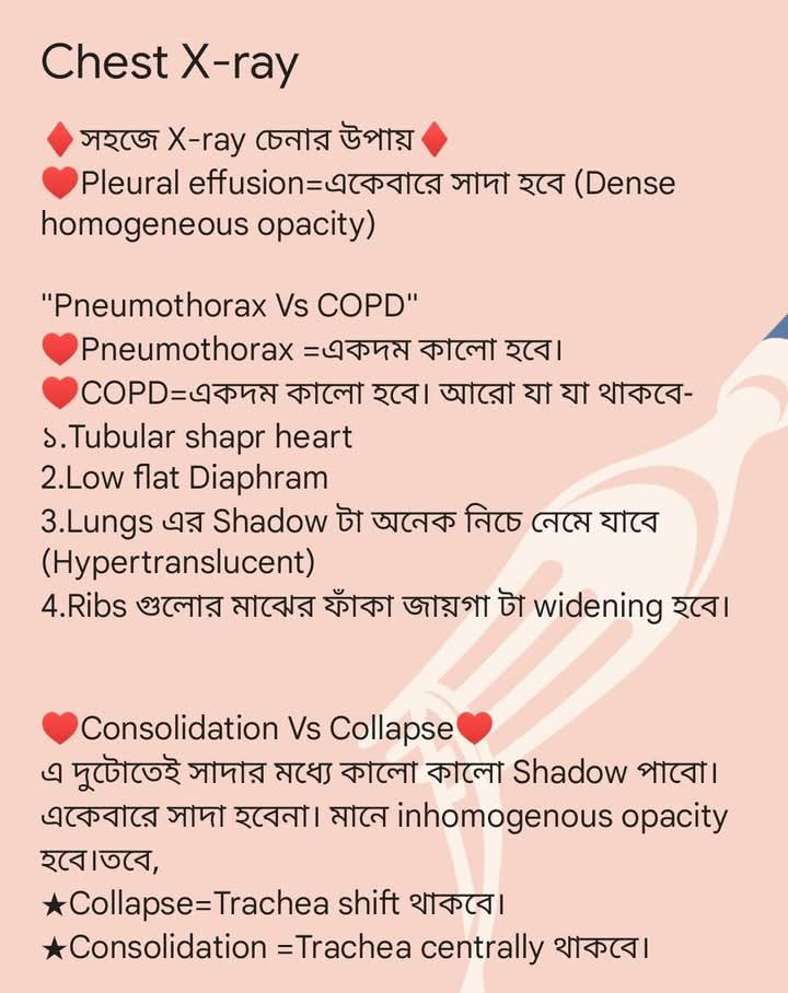
১. Pleural Effusion (Plural Fluid জমা হওয়া)
- X-ray Finding:
- একেবারে dense homogeneous সাদা opacity দেখা যাবে।
- সাধারণত নিচের দিক থেকে শুরু করে উপরে উঠবে (meniscus sign)।
- Fluid এর কারণে costophrenic angle blunt হয়ে যায়।
- Mediastinum/Trachea → opposite দিকে shift হতে পারে (বড় effusion এ)।
- Key clue: একেবারে সমান সাদা ছায়া (white out)।
২. Pneumothorax (Air in Pleural Space)
- X-ray Finding:
- একেবারে কালো (hyperlucent) area থাকবে, যেখানে কোনো vascular marking দেখা যাবে না।
- Lung collapse হয়ে পাশে চেপে থাকবে।
- Trachea shift → opposite দিকে (tension pneumothorax এ)।
- Key clue: খুব কালো এলাকা, lung margin দেখা যায়।
৩. COPD (Chronic Obstructive Pulmonary Disease)
- X-ray Findings:
- Lung fields দেখতে কালো (hypertranslucent), কিন্তু vascular markings থাকে।
- Tubular shaped heart (narrow vertical heart)।
- Diaphragm flat and low হয়ে যায়।
- Increased retrosternal air space (lateral view এ)।
- Rib spaces wide হয়ে যায় (barrel chest)।
- Key clue: Hyperinflation + flat diaphragm + tubular heart।
৪. Consolidation (Lung tissue filled with pus/blood/secretions)
- X-ray Findings:
- Homogeneous opacity (white patch) → কিন্তু এর মধ্যে air bronchogram দেখা যায় (air-filled bronchi inside opaque lung)।
- Trachea → usually central থাকে (shift হয় না)।
- Key clue: White opacity + air bronchogram + no tracheal shift।
৫. Collapse (Lung collapse / Atelectasis)
- X-ray Findings:
- White opacity → কিন্তু volume loss থাকবে।
- Trachea এবং mediastinum → opacity এর দিকে shift হবে।
- Adjacent ribs closer হয়ে যায়।
- Key clue: White opacity + trachea pulled towards lesion।
সহজে মনে রাখার Shortcut (Davidson অনুযায়ী):
- Pleural Effusion → সব সাদা + trachea opposite দিকে।
- Pneumothorax → সব কালো + lung margin দেখা যায় + vascular marking নেই।
- COPD → কালো lung field + flat diaphragm + tubular heart।
- Consolidation → সাদা patch + air bronchogram + trachea central।
- Collapse → সাদা patch + trachea একই দিকে টান পড়ে।
দারুণ 👍 এখন আমি Davidson অনুযায়ী Chest X-ray Findings এর Comparison Table বানিয়ে দিলাম। এক নজরে দেখে খুব সহজে পার্থক্য করতে পারবেন —
Chest X-ray Differential Table
| রোগ / অবস্থা | X-ray এ প্রধান Findings | Trachea / Mediastinum | Key Identification |
| Pleural Effusion | Dense homogeneous সাদা opacity; meniscus sign; costophrenic angle blunt | বড় হলে opposite দিকে shift | সব সাদা + fluid level |
| Pneumothorax | একেবারে কালো এলাকা (no vascular marking); lung margin দেখা যায় | Tension হলে opposite দিকে shift | Hyperlucent, lung margin visible |
| COPD | Hyperlucent lung fields; flat diaphragm; tubular heart; widened rib spaces | Usually central | কালো lung field + flat diaphragm |
| Consolidation | সাদা opacity + air bronchogram; homogeneous shadow | Trachea usually central থাকে | White opacity + air bronchogram |
| Collapse (Atelectasis) | সাদা opacity + volume loss; adjacent rib closer | Trachea same side এ shift | White opacity + tracheal pull |
✅ এভাবে মনে রাখবেনঃ
- সব সাদা হলে → Fluid (Effusion) বা Lung opacification (Consolidation/Collapse)
- সব কালো হলে → Air (Pneumothorax, COPD)
- Trachea opposite এ গেলে → Effusion / Tension Pneumothorax
- Trachea same side এ গেলে → Collapse
- Air bronchogram থাকলে → Consolidation
Raiyyan Tahsan Date:-20-08-2025 Wednesday Time:-02-11-25 Pm