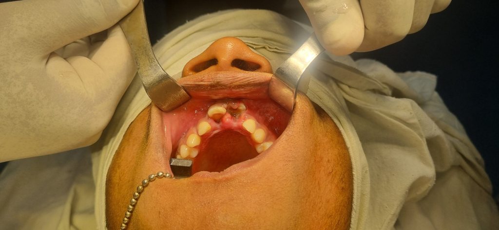
Management of intrusion of Maxillary Central incisiors due to trauma done by surgical repositioned & splinted by Arch bar wiring & lite cure composite filling under LA at OT complex of Oral and Maxillofacial department of Sir Salimullah Medical College and Mitford hospital, Dhaka.
The management of intrusion of maxillary central incisors due to trauma depends on several factors, including the severity of intrusion, the stage of root development, and the patient’s age. Here’s a step-by-step approach to managing this condition:
1. Clinical and Radiographic Assessment
Clinical Examination: Evaluate the degree of intrusion, mobility, and occlusion. Check for soft tissue injuries.
Radiographs: Take periapical and occlusal X-rays or OPG or CBCT to determine the extent of intrusion and root damage.
Pulp Testing: Assess pulp vitality, though it may not be reliable immediately after trauma.
2. Management Based on Severity of Intrusion
A. Mild Intrusion (≤3 mm)
Treatment: Allow spontaneous reeruption, especially in young patients with immature roots.
Follow-up: Monitor for signs of reeruption and pulp necrosis. If no movement occurs within 2–4 weeks, consider orthodontic extrusion.
B. Moderate Intrusion (3–6 mm)
Treatment: Orthodontic or surgical repositioning.
Orthodontic Repositioning: Brackets and very light forces applied over 5-6 months according to need.
Surgical Repositioning: Used if orthodontic extrusion is not feasible. The tooth is manually repositioned and stabilized with a flexible splint for 4 weeks.
Endodontic Considerations: If root development is complete, initiate root canal treatment (RCT) within 2–3 weeks to prevent pulp necrosis.
C. Severe Intrusion (>6 mm)
Treatment: Surgical repositioning is often required.
Splinting: A flexible splint for 4 weeks.
Endodontic Treatment: Immediate RCT in mature teeth; for immature teeth, monitor for pulp revascularization or perform apexification if necrosis occurs.
3. Post-Treatment Care
Splint Removal: After 4 weeks.
Pulp Vitality Testing: Regular checks at 2, 4, 6, and 12 months.
Radiographic Follow-up: Every 3–6 months for the first year, then annually for 5 years.
Monitor for Complications:
Pulp Necrosis: Indicated by discoloration or periapical radiolucency → requires RCT.
Root Resorption: External or replacement resorption → may need intervention.
Early intervention and regular monitoring are key to ensuring the best possible outcome.
Raiyyan Tahsan Date:-20-05-2025 Tuesday Time:-10-12-39 Pm